2D Electrophoresis
20 likes | 47 Vues
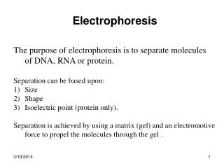
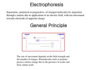
Creative Proteomics offers an integrated solution to the identification of low abundance proteins in complex biological sample through 2D electrophoresis. This technique sorts proteins according to two independent properties in two discrete steps. Each spot on the resulting two-dimensional array corresponds to a single protein species in the sample. Thus thousands of different proteins can be separated, and the information is obtained including the protein pI, the apparent molecular weight, and the amount of each protein.

2D Electrophoresis
E N D
Presentation Transcript
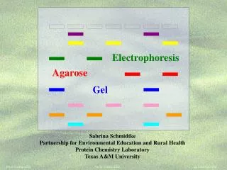
2-DE Creative Proteomics offers an integrated solution to the identification of low abundance proteins in complex biological sample through 2D electrophoresis. This technique sorts proteins according to two independent properties in two discrete steps. Each spot on the resulting two-dimensional array corresponds to a single protein species in the sample. Thus thousands of different proteins can be separated, and the information is obtained including the protein pI, the apparent molecular weight, and the amount of each protein. What Is 2D Electrophoresis? Two-dimensional gel electrophoresis, abbreviated as 2-DE, is a form of gel electrophoresis commonly used to analyze mixtures of proteins by two properties in two dimensions respectively. electrophoresis, proteins are first separated by their pI in isoelectric focusing and then further separated by molecular weight through SDS-PAGE, thus the sample proteins are distributed across the two-dimensional gel profile. This technique expands the number of proteins that can be identified, and provides more efficient data and detailed information for proteomics analysis. In two-dimensional gel 2D Electrophoresis 2-D electrophoresis begins with 1-D electrophoresis and then separates the molecules by a second property in a direction 90 degrees from the first. In 1-D electrophoresis, the proteins separated in one dimension will lie along a lane, and then the molecules are spread out across in the 2-D gel. Generally, it is unlikely that two molecules will be similar in both two distinct properties, so molecules are more effectively separated in 2-D electrophoresis than in 1-D electrophoresis. 2D Electrophoresis Service at Creative Proteomics 2-DE is a powerful and widely used method for the analysis of complex protein mixtures extracted from cells, tissues, or other biological samples. At Creative Proteomics, we can provide services 2D electrophoresis service according to standard operating procedures (SOPs). 2 DE. Before separating the proteins by mass in the second dimension, they are treated with sodium dodecyl sulfate (SDS) along with reducing reagents which unfolds them into long, straight molecules bound with a number of SDS molecules. Since the SDS molecules are negatively charged, all of the proteins
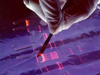
will have approximately the same mass-to-charge ratio as each other. In the second dimension, an electric potential is applied at a 90 degree angle from the first field. The gel therefore acts like a molecular sieve when the current is applied, separating the proteins on the basis of their molecular weight. The larger proteins remain higher in the gel and smaller proteins are able to pass through the sieve and reach lower regions of the gel. Protein detection. After 2-DE analysis, a gel with proteins spread out based on their pI and molecular mass is obtained. These proteins can then be detected by a variety of means, but the most commonly used stains are silver and coomassie brilliant blue staining. In the former case, a silver colloid is applied to the gel. The silver binds to cysteine groups within the protein and is darkened by exposure to ultra-violet light. The amount of silver relates to the darkness, and therefore the amount of protein at a given location on the gel. This measurement can only give approximate amounts, but is adequate for most purposes. Other staining methods such as coomassie brilliant blue combined with in-gel digestion are suitable for MS detection afterwards. Our Advantages The 2D SDS PAGE method has been standardized at Creative Proteomics through written SOPs. Proteins can be detected by various methods with the most commonly used stains (like silver and Coomassie Brilliant Blue staining) included to meet different requirements. Various computer-based methods are available for the detection and quantification of protein spots, such as SameSpots, Delta2D, ImageMaster, and so on. Based on professional knowledge and experienced staff, Creative Proteomics provides high-resolution 2-DE for separating all kinds of proteins. Our ordering procedure is as follows. As every project has different requirements, please contact our specialists to discuss your specific needs. If you have any questions or specific needs, don’t hesitate to contact us.