AMINO ACIDS
AMINO ACIDS. Proteins - biopolymers constructed from amino acids connected to each other by the peptide bonds. “ Protein ” - from the Greek word proteios , meaning primary or first rank. Proteins : • most abundant macromolecules in living systems;

AMINO ACIDS
E N D
Presentation Transcript
Proteins - biopolymers constructed from amino acids connected to each other by the peptide bonds “Protein” - from the Greek word proteios, meaning primary or first rank Proteins: • most abundant macromolecules in living systems; • occur in great variety, most diverse macromolecules in living systems; • function in all biological processes
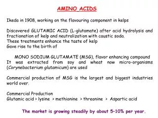
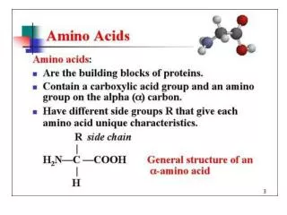
Amino acids - building blocks of proteins Amino acid- any organic molecule with at least one carboxyl group and at least one amino group Several hundreds of different amino acids are known In animal cells there are 20main amino acids that are genetically coded for incorporation into protein
20 amino acids found in proteins 3 letter abbreviation or 1 letter symbol are used to indicate the composition and sequence of amino acids in proteins
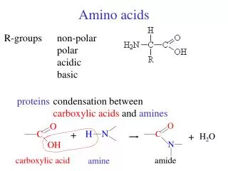
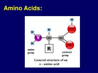
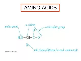
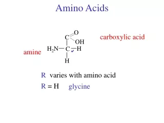
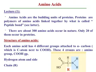
side chain General formula of amino acid Amino acid has a central tetrahedral carbon atom (α-carbon) linked to: - an amino group - a carboxylic acid group - a hydrogen atom - side chain (R group) Because of functional groups are placed around the α-carbon center, they are called α-amino acids α-Amino acids are chiral - 4 different groupsconnected to the tetrahedral α-carbon (except for glycine which is achiral)
Classification of amino acids The feature making each amino acid chemically and biologically distinct is theR side chain • 20 side chains are varied in: • size, • shape, • charge, • H-bonding capacity, • hydrophobic character, • chemical reactivity All amino acids are divided into classes based on polarity of the side chain
Group 1. Amino acids with nonpolar side chain Glycin, alanin, valine, leucine, isoleucine, proline, phenylalanine, methyonine, tryptophan All amino acids of this group have aliphatic or aromatic groups and therefore have hydrophobic character
Cysteine (Cys, C) Serine (Ser, S) Threonine (Thr, T) Group 2. Amino acids with polar, uncharged side chains Serine, cysteine, threonine, tyrosine, asparagine, glutamine Side chains of these amino acids have heteroatom (N, O or S) with electron pair available for hydrogen bonding to water and other molecules
Asparagine (Asn, N) Glutamine (Gln, Q)
Group 3. Amino acids with polar, negatively charged side chains at physiological pH Glutamate, aspartate Contain acidic functional groups At physiological pH the side chain dissociate protons to form carboxylate anions Aspartate (Asp, D) Glutamate (Glu, E)
Group 4. Amino acids with polar, positively charged side chains at physiological pH Lysine, arginine, histidine Contain basic functional groups Lysine (Lys, K) Arginine (Arg, R)
Stereochemistry of Amino Acids Amino acids exist in two stereoisomers (enantiomers), called D- and L-aminoacids D- and L-isomers are mirror image of each other Only the L-isomers are used as building blocks of proteins D-isomer L-isomer
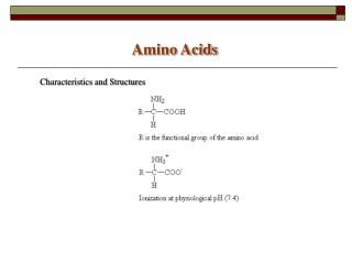
Properties of amino acids In pure form are wide,crystalline, high-melting solids Soluble in water and insoluble in organic solvents Amphoteric properties – depending on medium can act as acids or basis With base amino acids react as acids: With acids amino acids react as bases:
Amino acids are ionized in aqueous solutions At physiological pH amino acids have a positive and negative charge on the same molecule (zwitterions) The net charge of the zwitterion form is 0 Zwitterions– the fully ionized species of amino acids having one amino group and one carboxyl group
Ionization of amino acids At high pH, the protonated amino group loses a proton and amino acid has negative charge At low pH (acid solution) the dissociated carboxyl group accept the proton and amino acid has positive charge
Proteins - biopolymers constructed from amino acids connected to each other by the peptide bonds FUNCTIONS OF PROTEINS • Enzymes (catalysts of biochemical reactions) All enzymes of living cells are simple or complex proteins • Structural function Fibrous proteins collagen and elastin are the constituents of bones, skin, tendons, cartilage, walls of vessels Keratin – main component of hair and nails Proteins of cell membranes • Immune function Immunoglobulins(antibodies) are produced in response to the antigen (bacteria, foreign proteins) invasion into the organism • Transport function Hemoglobin – transport of oxygen and carbon dioxide Lipoproteins – transport of cholesterol, triacylglycerol via the blood
Storage function Store nutrients for future use Casein – protein of milk Myoglobin – depot of oxygen in muscles Proteins of blood plasma (albumins and globulins) store amino acids • Regulatory function Many hormones are proteins • Receptor function Receptors are located on cell membranes or in the cytoplasm and play role in the signal generation and transmission • Muscle contraction Actin, myosin, troponin, tropomyosin – functional components of the contractile system of skeletal muscles
Levels of protein structure • Primary • Secondary • Tertiary • Quaternary
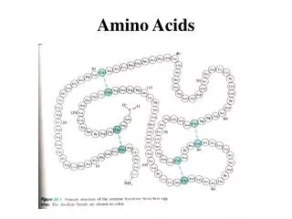
Primary structure of the proteins Primary structure- type and sequence of amino acids in a polypeptide chain Example: -Met–Glu–Asp–Phe–Gly–Leu–Ala-Met–Glu–Gly– Amino acid type and sequence is determined genetically Primary structure is formed due to the formation of peptide bonds between amino acids
Peptide bond - amide bond that is formed between α-carboxyl group and α-amino group of two amino acids During the formation of peptide bond the molecule of H2O is released Peptide bond is a covalent, stable bond Dipeptide - two amino acids are linked together Tripeptide - three amino acids… Polypeptides – many amino acids…
Polypeptides have polarity Amino and carboxyl groups of amino acids are used for peptide bond formation There are free amino and free carboxyl group at the ends of polypeptide chain The amino group end is called N-terminal residue and the end containing free carboxyl group is called C-terminal residue
Polypeptide chain consist of regularly repeating part, so called backbone, and side chains which are variable in different amino acids In the polypeptide chain the carboxyl groups and amino groups are involved in peptide bond formation and can not dissociate and are not functionally active The properties of polypeptide are determined by the side chains of the amino acid
Structure of the peptide bond Rotation about the amide carbon nitrogen (peptide) bond is limited Four atoms of the peptide group are on the same plane Peptide bonds are rigid Peptide group is in trans configuration: hydrogen atom of the nitrogen is opposite to the oxygen atom of the carbonyl group Peptide group acts as single, planar unit
Naming of peptides Peptides are named as acyl derivatives (the –ine ending is changed to –yl) of amino acids listed in the order in which they appear, starting with the N-terminal amino acid Alanyltyrosylglycine
Determination of primary structure of polypeptides Sanger’s method Sanger’s reagent (2,4-dinitrofluorobenzene (DNFB)) reacts with amino group of the N-terminal amino acid of a polipeptide chain The carbon-nitrogen bond between amino acid and DNFB is more resistant to hydrolysis than the peptide bonds Substituted polipeptide is hydrolized Terminal amino acid remains with the DNFB and can be isolated and identified
Edman’s method Allows to label N-terminal residue and cleave it without hydrolyzing other amino acids Steps: 1. Phenyl isothiocyanate react with the amino group of N-terminal residue 2. Releasing of the modified amino acid (cyclic derivative) 3. Identifying of the modified amino acid by chromatographic procedures (HPLC) 4. Repeating on next amino acid….
Secondary structure of protein molecule • spatial conformation of the protein backbone • spatial arrangement of amino acids that are nearby in sequence Secondary structure - regularly repeating conformations of the peptide chain, such as α-helices and β-sheets The two most important types of secondary structure: -α-helix -β-pleated sheet
The interaction that hold the polypeptide unit in the arrangement are hydrogen bonds between N-H group of one anino acid residue and C O group of the amino acid four residues ahead in the chain α-helix α-helix - a rodlike structure formed by tightly coiled polypeptide backbone Helix is stabilized by many hydrogen bonds (which are parallel to long axis of the helix)
Most a helices in proteins are right handed (backbone turns clockwise when viewed along the axis from the N terminus) N Hydrogen bonds allow the polypeptide to stretch up to twice its normal length The R-side chains branch out from the main chain Right-handed (clockwise) rod structure O
b-Sheets Inbsheet configuration polypeptide chain form an extended zigzag Side chains alternate above and below plane Multiple polypeptide chains can be arranged side-by-side Polypeptide chains in -sheet are stabilized by hydrogen bonds between C=O and NH on adjacent chains
Tertiary Structure of Proteins • folding of a polypeptide chain into a closely-packed three-dimensional structure • the spatial arrangement of amino acids that are far apart in the sequence (amino acids far apart in the primary structure may be brought together) • stabilizedby noncovalent interactions (e.g. hydrophobic effects, charge-charge interaction) between side chains or disulfide bridges
Quaternary Structure Monomeric proteins - consist of only one polypeptide chain Oligomeric proteins – consist of two or more polypeptide chain called subunits Quaternary structure – arrangement and position of each subunits in the intact protein molecule (3D structure of 2 or more polypeptide chains) Subunits are held together by many weak, noncovalent interactions (hydrophobic, electrostatic)
Levels of protein structure •Primary •Secondary •Tertiary •Quaternary
Three dimensional conformations of secondary, tertiary and quaternary structures are held together by a variety of interactions (bonds) between amino acid side chains Bonds maintaining spatial conformations of proteins: -disulfide bonds -hydrogen bonds -charge-charge forces (ionic bonds) -hydrophobic forces Disulfide bond Disulfide bond maintains the secondary, tertiary and quaternary structures of proteins It can be formed between two cysteines of the same polypeptide chain or adjacent chains
Disulfide bonds Formation of disulfide bond Disulfide bond– bond between two sulfur atom of amino acid cysteine It is formed by the oxidation of a pair of cysteine residues
Hydrogen bonds in secondary structure occur betweenN-H group of one anino acid residue and C O group of the amino acid four residues ahead in the chain Hydrogen bonds Stabilizes secondary, tertiary and quaternary structures Helix is stabilized by many hydrogen bonds which are parallel to long axis of the helix
Charge-charge interaction (ionic bonds) • interaction between two charged amino acids • negatively charged amino acids: glutamate and aspartate; • positively charged amino acids: lysine, arginine and hystidine
CH3 The hydrophobic effect Nonpolar side chains of amino acids associate with each other Nonpolar side chains are in the interior Nonpolar molecules are aggregated because polar water molecules push away nonpolar compounds (nonpolar compounds want to escape from water)
Loss of Protein Structure (Denaturation and Hydrolysis) • Denaturation - disruption of native conformation (loss of organized structure) of a protein, with loss of biological activity • The stabilizing forces holding a protein in its native conformation are relatively weak so they can be disrupted under mild conditions • During denaturation only higher structures (secondary, tertiary and quaternary) of proteins are disrupted. Primary structure remains unchanged • Denaturation is irreversible process • Some proteins can be refolded (renatured)
Denaturants • Heat (example: denaturation of egg white) t > 50oC breaks H-bonds • Strong acids and alkalines • Organic solvents (ethyl alcohol, acetone) • Detergents • Agents reducing disulfide bonds (-mercaptoethanol) • Agents disrupting H-bonds (urea)
The tertiary structure of ribonuclease is maintained by four S-S bridges Denaturation of ribonuclease with urea and mercaptoethanol results in complete loss of tertiary structure and enzymatic activity and yields a polipeptide chain containing eight sulfhydryl groups
Hydrolysis – breaking of the peptide bonds with free amino acid formation During hydrolysis the primary structure of proteins is destroyed Acid and alkaline hydrolysis – boiling of proteins in the presence of strong acids or bases Enzymatic hydrolysis takes place in the stomach and intestine (dietary proteins are hydrolyzed by the enzymes peptidases)
Examples of proteins and their structure Fibrous Proteins • have “fiber-like” or elongated shape • have highly developed secondary structures • Keratin is found in hair, feathers, horns (depends on the –helix for strength) • Fibroin is folded into –sheet secondary structure. It is found in silk • Collagen is a major protein in connective tissue of vertebrates (25-35% of total protein in mammals) • - forms the bone matrix, ligaments, tendons and skin • - consists of three helical chains coiled around each other in supercoil • - is extremely strong and stable
Keratin, fibroin and collagen – examples of fibrous proteins