Download
1 / 112
1.13k likes | 1.42k Vues
Nerve and Muscle. Physiology of nerve. The neuron. The basic structural unit of the nervous system. Structure: The soma The dendrites: antenna like processes The axon: hillock, terminal buttons. Types of nerve fibers. a- myelinated nerve fiber:

E N D
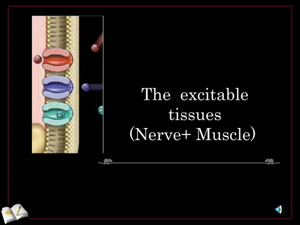
Nerve and Muscle Physiology of nerve
The neuron • The basic structural unit of the nervous system. • Structure: • The soma • The dendrites: antenna like processes • The axon: hillock, terminal buttons
Types of nerve fibers a- myelinated nerve fiber: • Covered by myelin sheath, protein-lipid layer, secreted by Schwann cells, acts as insulator to ion flow, interrupted at Nodes of Ranvier b- unmyelinated nerve fiber: • Less than 1μ, covered only with Schwann cells, as postganglionic fibers
Electrical properties of a neuron • Electrical properties of nerve & muscle are: • 1-There is difference in electrical potential between the inside and outside the membrane • 2-Excitability: the ability to respond to any stimulus by generating action potential • 3-Conductivity: the ability to propagate action potential from point of generation to resting point
Membrane potential; the basis of excitability • Def:electrical difference between the inside & outside the cell • Causes:selective permeability of the membrane • more K+, Mg2+, Ptn, PO4 inside • more Na+, Cl-, HCO3-outside • Exists in all living cells & it is the basis of excitability • Excitability: • Def: it is the ability to respond to stimuli (change in the environment) giving a response • The most excitable tissues are nerves & muscles • Stimuli: • Types: • Electrical (preferred), chemical, mechanical, or thermal. • Cathode ( more important) & anode +anode - cathode
Excitability • Factors affecting effectiveness of the stimulus: • 1- strength: • effective stimulus • 2- duration: • a certain period of time, very short duration can not excite the nerve • 3- rate of rise of stimulus intensity: • Rapid increase…. Active response • Slow increase …. adaptation
Strength –Duration Curve • Within limits stronger intensity shorter duration • Strength: • Threshold stimulus (rheobase): it is the minimal amplitude of stimulus that can excite the nerve and produce action potential. • Subthreshold stimulus: causes local response (electrotonic) • Duration: • stimuli of very short duration can not excite the nerve • Utilization time: is the time needed by threshold stimulus (Rheobase) to give a response • :Chronaxietime needed by a stimulus double the rheobaseto excite the nerve, it is a measure of excitability, decrease chronaxie means increase excitability
Strength –Duration Curve Stimulus amplitude chronaxie 2R Utilization time R duration
Measuring the membrane potential Recording: by 2 micoelectrodes inserting one inside the fiber & the other on the surface & connected to a voltmeter through an amplifier
Types of membrane potential • Membrane potential has many forms: • 1- RMP • 2- on stimulation; • a) action potential if threshold stimulus • b) localized response (electrotonic) if subthreshold stimulus
Resting membrane potential (RMP) *definition: It is the difference in electrical potential between the inside and outside the cell membrane under resting conditions with the inside negative to the outside • Value:-90 mv large fibers, -70 in medium fibers, -20 in RBCs • Causes • 1- selective permeability • 2- Na-K pump Recording: by 2 micoelectrodes inserting one inside the fiber & the other on the surface & connected to a voltmeter
Resting Membrane Potential Selective permeability of the membrane: contributes to -86mv • K+, ptn-, Mg2+&PO4- are concentrated inside the cell • Na+, Cl-, HCO3- are found in the extracellular fluid • During rest the membrane is 100 times more permeable to K+ than to Na+, • K+tend to move outward through INWARD RECTFIER K+ channels down their concentration gradient • The membrane is impermeable to intracellular Ptn-&other organic ions • Accumulation of +ve charges outside & -ve charges in • At equilibrium :K+ in to out is 35:1 • Na+ in to out is 1-10
Potassium equilibrium -90 mV
Na-K pump • Definition: carrier protein on the cell membrane: • 3 binding sites inside for Na+ • 2 sites outside for K+ • 1 site for ATP • Inner part has ATPase activity • It is an electrogenic pump Contributes for -4mv and helps to keep RMP
Nernest equation • E for K = -61 log con inside/ conc outside =- 94 • E for Na = -61 log con inside/ conc outside =+ 61 • Goldman equation: it considers • 1- Na, K and cl concentrations. • 2- K permeability is 100 times as that for Na
Action Potential • Definition: It is the rapid change in membrane potential following stimulation of the nerve by a threshold stimulus. • Recording: microelectrodes and oscilloscope.
Membrane Permeabilites • AP is produced by an increase in Na+ permeability. • After short delay, increase in K+ permeability. Figure 7-14
Shape and Phases of Action Potential • 1- Stimulus artifact.: small deflection indicates the time of application of stimulus, it is due to leakage of current • 2- Latent Period: isoelectrical interval, time for AP to travel from site of stimulation to recording electrode. • 3- Ascending limb (depolarization):starts slowly from -90, till firing level-65mv, reaches &overshoots the isopotential, ends at +35 • 4- Descending limb:(repolarization): starts rapidly till 70% complete then slows down * Hyperpolarization: in the opposite direction slight & prolonged • 5- RMP
Shape and Phases of Action Potential 1- Ascending limb (depolarization) Slow..firing level..rapid. 2- Descending limb (repolarization) rapid then slow 3- Hyperpolarization: slight & prolonged 4- RMP +35 0 -65 -90 overshoot repolarization depolarization mv FL hyperpolarization Latent period time
Duration of Action Potential • Spike lasts 2msec • Hyperpolarization 35-40msec
Ionic basis of action potential • Depolarization is caused by Na+ inflow • Repolarization is caused by K+ outflow Two types of gates: 1- voltage gated Na+ channels; having 2 gates: outer activation gate & inner inactivation gate 2- voltage gated K+ channels; one activation gate When the nerve is stimulated:: a- the outer gate of VG Na+ opens, activating Na+ channel…. Na+ inflow b- the inner gate of Na+ channels closes, inactivating Na+ channels… stop Na inflow c- K+ gates open, activating K+ channels, K+ outflow
The Action Potential A stimulus opens activation gate of some Na+ channels depolarizing membrane potential, allowing some Na to enter, causing further depolariztion If threshold potential is reached, all Na+ channels open, triggering an action potential.
The Action Potential 1-Depolariztaion:occurs in 2 stages: Slow stage: -90 to -65mv: some Na+ channels opened, depolarizing membrane potential, allowing some Na to enter, causing further depolarization At -65mv, the firing level or threshold for stimulation, all Na+ channels open, triggering an action potential. Rapid stage: -65 to +35: all Na+ channels are opened, Na+ rush into the fiber, causing rapid depolarization
The Action Potential Within a fraction of msec, Na+ channel inactivationgates close and remained in the closed state for few milliseconds, before returning to the resting state. 2- Repolarization: Inactivation of Na+ channels and activation of K+ channels are fully open. Efflux of K+ from the cell drops membrane potential back to and below resting potential 3- Hyperpolarization; slow closure of K+ channels
The Action Potential The Na+ & K+ gradients after action potential are re-established by Na+/K+ pump Only very minute fraction of Na+ & K+ share in action potential from the total concentration The action potential is an all-or-none response. (provided that all conditions are constant, AP once produced, is of maximum amplitude, constant duration & form, regardless the amplitude of the stimulus , however threshold or above Action potential will not occur unless depolarization reaches the FL (none) Action potential size is independent of the stimulus and once depolarization reaches FL, maximum response is produced, reaches a value of about +35 mV(all)
The Action Potential Both gates of Na+ channel are closed but K+ channels are still open. Continued efflux of K+ keeps potential below resting level. K+ channels finally close and Na+ channel inactivation gates open to return to resting state.
Action potential initiation S.I.Z.
Action potential in a nerve trunk • Nerve trunk is made of many nerve fibers • The AP recorded is compound action potential, having many peaks • The individual fibers vary in: • 1- threshold of stimulation • 2-distance from stimulating electrode • 3- speed of conduction
During depolarization, there is +ve feed back response. • Repolarization is due to: 1- inactivation of Na+ channels( must be removed before another AP 2- slower & more prolonged activation of K+ channels • Hyperpolarization (undershoot): slow closing of K+ channels, K+ conductance is more than in resting states • Role of Inward rectifier K+ channels: Non gated channels Tend to drive the membrane to the RMP Drive K+ inwards only in hyperpolarization Re-establishing Na+ &K+ gradient after AP:role of Na+ /K+ pump All or none law
Electrotonic potentials & local response • Catelectronus: at cathode/ depolarization less than 7mV/ passive • Anelectronus: at anode/ hyperpolarization/ passive • Local response (local excitatory state): • Stonger cathodal stimuli • Slight active response • Some Na+ channels open, not enough to reach FL • It is graded • Does not obey all or none law • Non propagated • Excitability of the nerve increased • Caused by subthreshold stimulus • Can be summated & produce AP • Has no refractory period
Local Response (local excitatory change) • Although subthreshold stimuli do not produce AP they produce slight active changes in the membrane that DO NOT PROPAGATE. • It is a state of slight depolarization caused by subthreshold cathodal stimulus that opens a few Na channelsnot enough to produce AP
Local Response (local excitatory change) • It differs from AP : • It does not obey all or non rule • Can be graded. • Can be summated. • It does not propagate.
Excitability changes during the action potential • Up to FL, excitability increases The remaining part of action potential, the nerve is refractory to stimulation (difficult to be restimulated) • Absolute refractory period: Def: the period during which a 2nd AP can not be produced whatever the strength of the stimulus Length: from FL to early part of repolarization Causes: inactivation of Na+ channels • Relative refractory period: Def.; the period during which membrane can produce another action potential, but requires stronger stimulus. Length: from after the ARP to the end of the AP Causes: some Na+ channels are still inactivated K+ channels are wide open. ARP RRP FL Increased excitability
Factors affecting Membane potential & Excitability • Factors ↑ excitability: • * Role of Na+ • 1) ↑ Na permeability (veratrine & low Ca 2+). • Factors ↓ excitability: • 1)↓ Na permeability( local anaesthesia & high Ca2+) [ membrane stbilizers] • Decrease Na+ in ECF: decreases size of AP, not affecting RMP • Blockade of Na+ channels by tetradotoxin TTX decrease excitability & no AP • ** Role of K+: • 1)↑ K extracellularly (hyperkalemia). • 2)↓ K extracellularly (hypokalemia): familial periodic paralysis • 3) blockade of K+ channels by TEA: prolonged repolarization& absent hyperpolarization • *** Role of Na+ K+ pump: only prolonged blockade can affect RMP & AP
Accommodation of nerve fiber • Slow increase in the stimulus intensity gives no response: • 1- inactivation of Na+ Channels • 2- opening of K+ Channels
Conduction in an Unmyelinated Axon • The action potential generated at one site, acts as a stimulus on the adjacent regions • During reversal of polarity, the stimulated area acts as a current sink for the adjacent area • A local circuit of current flow occurs between depolarized segment & resting segments (flow of +ve charges) in a complete loop of current flow • The adjacent segments become depolarized, FL is reached, AP is generated Figure 7-18
Conduction in Myelinated Axon (Saltatory conduction) • Myelin prevents movement of Na+ and K+ through the membrane. • The conduction is the same in unmyelinated nerve fibers Except that AP is generated only at Nodes of Ranvier • AP occurs only at the nodes. • AP at 1 node depolarizes membrane to reach threshold at next node. • The +ve charges jump from resting Node to the the neighbouring activated one (Saltatory conduction). Figure 7-19
Importance of saltatory conduction: • ↑velocity of nerve conduction. • Conserve energy for the axon.
Orthodromic & antidromic conduction • Orthodromic: from axon to its termination • Antidromic: in the opposite direction • Any antidromic impulse produced, it fails to pass the 1st synapse & die out
Monophasic &biphasic AP • Monophasic AP: recorded by one microelectrode inserted inside the fiber & one indifferent microelectrode on the surface. • Biphasic: two recording electrodes on the outer connected to CRO
Depolarization & repolarization of a nerve fiber • RMP does not record any change • Depolarization flows to the +ve electrode ..... Upright deflection (+ve wave) • Complete depolarization ... No flow of current (baseline) • Repolarization to the +ve electrode....down deflection • Complete repolarization ... No flow of current (baseline)
Action potential in a nerve trunk • Nerve trunk is made of many nerve fibers • The AP recorded is compound action potential, having many peaks • The individual fibers vary in: • 1- threshold of stimulation • 2-distance from stimulating electrode • 3- speed of conduction
Compound AP • Graded • Subthreshold; no response occurs • Threshold; a small AP, few nerve fibers • Further increasing; AP amplitude increases up to a maximal • Increasing the intensity, supramaximal stimuli, no more increase in the AP
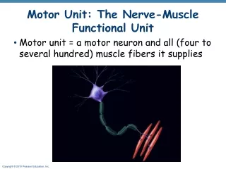
Nerve fiber types • According to their thickness, they are divided into:
Metabolism of the nerve • Rest: nerve needs energy to maintain polarization of the membrane, energy needed for Na+/K+ pump, derived from ATP. Resting heat • Activity: pump activity increases to the 3rd power of Na+ concentration inside, if Na+ concentration is doubled, the pump activity increases 8 folds;23 . • Heat production increases: • 1- initial heat during AP • 2- a recovery heat, follows activity =30 times the initial heat • Neurotrophins: • Proteins necessary for neuronal development, growth & survival • Secreted by glial cells, muscles or other structures that neuron innervate • Internalised & retrograde transported to the cell body
Types of muscles • Skeletal muscle: under voluntary control 40% of total body mass. • Cardiac muscle: not under voluntary control. • Smooth muscle: not under voluntary control. Both are 10% of total body mass
Skeletal muscles • Attached to bones • >400 voluntary skeletal muscles • Contraction depends on their nerve supply • 4 functions: • 1- force for locomotion & breathing • 2- force for maintaining posture & stabilizing joints • 3- heat production • 4- help venous return
Morphology • Muscle fibers: • Bundled together by C.T. • Arranged in parallel between 2 tendenious ends • Is a single cell • Closely enveloped by glycoprotein sheath (sarcolemma) outside the cell membrane • Made of many parallel myofibrilsembeded in a sarcoplasm, between a complex tubular system
Skeletal muscle • Each muscle fiber is a single unit. It is made up of many parallel myofibrilsembedded together and a complex sarcotubular system. • Each muscle fibril contains interdigitating thick and thin myofilaments arranged in sarcomeres. • 2 major proteins: • 1- thick filaments [myosin] • 2- thin filaments [actin, troponin, troopomyosin] • Troponin & trpomyosin regulate muscle contraction by controlling the interaction of actin & myosin