Download
1 / 14
150 likes | 347 Vues
DNA and Amino Acids. Molecular Structure Lecture 3. Chromosome to DNA molecule. A chromosome is essentially a long strand of DNA wound around proteins; e.g. histones , to form condensed structure called chromatin.

E N D
DNA and Amino Acids Molecular Structure Lecture 3
Chromosome to DNA molecule • A chromosome is essentially a long strand of DNA wound around proteins; e.g. histones, to form condensed structure called chromatin. • However it order for the DNA to carry out its function is must be unwound from the proteins: chromatin to a long strand of DNA • This DNA is shaped in the form of, the now famous, “double Helix” • DNA is an abbrevation for Deoxyribose Nucleic acid [see next slide] • It consists of a long strand of DNA nucleotides which are joined together. • The DNA “double Helix” is two such strands which are coiled and connected together via what are referred to as: nucleotide bases or bases
The Crick and Watson double Helix Adapted from [1] p.194 [the original article by C and W p 195]
RNA: Ribose nucleic acid • A molecule closely associate with DNA and which a part of the “gene expression” process is referred to as RNA • The RNA [nucleotide] is very similar to DNA [nucleotide] except: • Its nucleic acid has a ribose sugar as opposed to a deoxyribose sugar. • The Neuleotide base thymine is replace with an equivalent base called Uracil [klug p.191] • The RNA strand unlike the DNA double helix strand is single stranded
DNA version of genetic code table • Later we will see how gene expression and protein production [the basis of life] works. • The process involves the use of a genetic code where sets of 3, triplets, of nucletides bases [Codon] is converted into an amino acid [ described shortly] • The following tables show the code form the prespective of the DNA codon or RNA codon
The genetic code RNA conversion table DNA conversion table Note: the only difference is T being replaced with U Adapted from Ref [1] p. 247
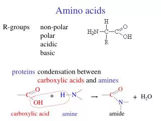
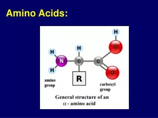
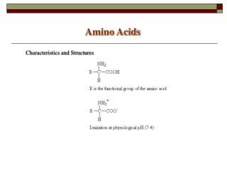
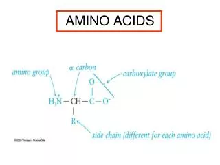
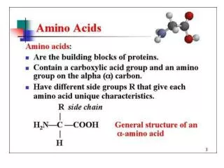
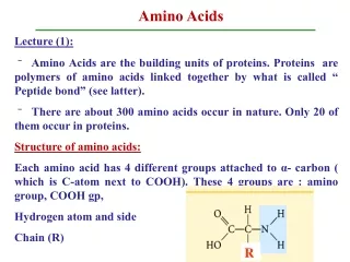
Amino acids • The genetic code table shows DNA/RNA being converted into “amino acids” • An amino is a molecule that has two main elements: a constant part [shown in pink in the next slide] and a variable part. This variable part has specific chemical properties which are essential to its function. • In proteins [chains of amino acids] the constant regions are referred to as the “backbone” and the variable region as the side chains.
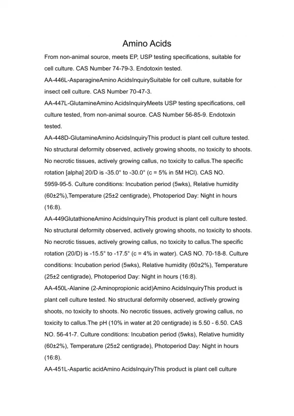
AMINO Acids (AA) and their properties Alanine-> name of Amino acid Ala -> the common abbreviation A -> code used in protein sequences (this letter is not always the first letter of the AA; e.g. tryptophan is W) MW: molecular weight or weight of the amino acid “the amino acids are grouped according to their polarity and charge. They are divided into four categories, those with polar uncharged R groups [hydrophilic], those with (nonpolar) R groups [hydrophobic], acidic and basic groups.” Ref [2]. Also Cystine is the only amino acid with a sulphur atom.
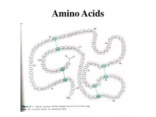
An amino chain or Polypeptide • When two AA are joined together it is by, what are called, “peptide” bonds of the constant element of each amino acid • A third is the joined to the second using the same type of connection or “bond” • When a number of AAs are joined together they start to form the main chain • Subsequent addition results in a polypeptide chainor(primary protein structure). the peptide has two elements: the main chain connected via peptide bonds; and side chains ( generally associated with functionality).
Polypeptide Bond • When two amino acids bond together a H20 water molecule is formed along with the peptide bond
Polypetide chain -> Secondary structure The primary chain (polypeptide chain) then begins to change its shape depending on the side chain properties of the amino acid to firstly form: the secondary structure: e.g. α helixes and β sheets both These structures are found in all proteins and form the next level in the formation of the 3D structure The pink “backbone” corresponds to main chain the other elements show side chains and their bonding
Secondary to Tertiary structure Secondary structures interact to form the tertiary or 3-D structure of the protein : essentially the polypeptide chain after it has been twisted and turned to form the most thermodynamically stable structure: Since most proteins are in water A number of factors affect the formation: Polar (Hydrophilic) AA try to stay on the outside of the structure Non polar (hydrophobic) stay on the inside disulphide bonds between cistine AA However in some cases the proteins are in hydrophobic solution [Lipids in the cell membrane] and in this case the structure would alter: non polar outside, polar inside. Myoglobin: Adapted from [1] p 279
Exam question • Discuss how a DNA sequence forms the final 3-D configuration of proteins. • Discuss why a DNA is crucial in the formation of a 3-D protein structure.
References • [1] Klug 7thed • [2] Http://biotech.matcmadison.edu/resources/proteins/labManual/chapter_2.htm; accessed on the 21/9/2011