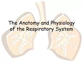
Respiratory System & Emergencies
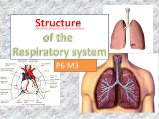
Respiratory System & Emergencies. Respiratory system. The respiratory system is the KEY component in patient care. If you have an airway or breathing problem nothing else matters... . Structure & Function. Respiratory system is divided into two parts: The upper airway & lower airway.

Respiratory System & Emergencies
E N D
Presentation Transcript
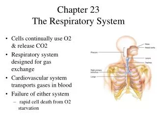
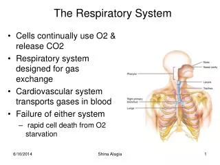
Respiratory system The respiratory system is the KEY component in patient care. If you have an airway or breathing problem nothing else matters...
Structure & Function Respiratory system is divided into two parts: The upper airway & lower airway
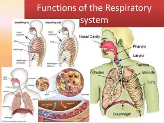
Upper airway • From nose/mouth to epiglottis. • Filters air • Warms & humidifies air • provides sense of smell
Lower airway • From epiglottis to alveoli • Contains Larynx (vocal cords) • Exchange of oxygen (O2) & carbon dioxide (CO2)
Pharynx • Oropharynx and Nasopharynx • location of gag reflex for airway protection
Trachea • conducts air into lungs • hard - cartilage
Bronchi • connects trachea to each lung • angle difference • straight into right lung • sharp angle into left lung
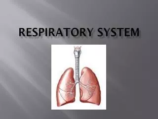
Lungs • organ of respiration where exchange of O2 & CO2 take place • Each lung divided into lobes • location during ... • inspiration - the umbilicus • expiration - 4-5 intercostal space
Alveoli • smallest unit of respiratory system • microscopic sacks surrounded by capillaries • Gas exchange
Pleura • Parietal pleura • Visceral pleura
Diaphragm • Has characteristics of both voluntary and involuntary muscles • Dome-shaped • Divides thorax from abdomen • Contracts during inhalation • Relaxes during exhalation
Protective Structures • rib cage - protects from trauma • cilia - removes microscopic particles • pleura – reduces friction
Breathing vs Respiration • NOT THE SAME • breathing = moves air in & out of system • respiration = supplies cells with O2 & removes CO2 - exchange at cellular level
Breathing Process: Inhalation • Diaphragm and intercostal muscles contract • Size of thoracic cavity increases • Pressure in the lungs decreases • Air travels to the lungs
Breathing Process: Exhalation • Diaphragm and intercostal muscles relax. • All dimensions of the thorax decrease. • Pressure in the lungs increases. • Air flows out of the lungs.
Exchange of Oxygen and Carbon Dioxide • Oxygen-rich air is delivered to alveoli. • Oxygen diffuses into the blood. • The body does not use all the inhaled oxygen.
Respiratory Center • located in brainstem • responds to CO2 • controls rate & tidal volume • phrenic nerve makes the diaphragm work (neck injury can injure this nerve C3-5)
Signs of Adequate Respirations • Effortless • NTV • Normal rate • Regular rhythm • Normal LOC • Warm skin with normal color • Normal breath sounds bilaterally
Signs of Respiratory Distress (1 of 2) • Slower than 8 breaths/min or faster than 24 breaths/min • Irregular rhythm • Quality - labored • Accessory muscle use • Noisy or diminished breath sounds
Signs of Respiratory Distress(2 of 2) • Pale or blue skin • Cool, clammy skin • Dyspnea • Conversational dyspnea • Tripod position • Anxiety
Infant and Child Considerations • Structures less rigid • Airway smaller • Larger tongue • More dependence on diaphragm • Nasal flaring and seesaw respirations
Respiratory Emergencies & Dyspnea
Dyspnea • Shortness of breath (SOB) or difficulty breathing • Patient may not be alert enough to complain of shortness of breath
Terminology • Hypoxia - decreased/low oxygen • Anoxia - no oxygen • Wheezing - whistling sound • Rales - fluid sound (crackles) • Stridor - high pitched sound • Rhonchi - coarse gravelly sounds (similar to wheezing)
Causes of Poor Breathing • Pulmonary vessels become obstructed. • Alveoli are damaged. • Air passages are obstructed. • Blood flow to the lungs is obstructed. • Pleural space is filled.
Abnormal Respiratory Patterns • Apnea • Cheyne-Stokes • Central Nervous System Hyperventilation* • Ataxic *Similar to Kussmal respirations in a diabetic patient
Upper or Lower Airway Infection • Infectious diseases may affect all parts of the airway. • Cause obstruction
Acute Pulmonary Edema • Fluid build-up in the lungs • Signs and symptoms • Dyspnea • Frothy pink sputum • Rales (crackles) • Recurrence high • History of chronic congestive heart failure
Chronic Obstructive Pulmonary Disease (COPD) • Emphysema or chronic bronchitis • Barrel chest • Labored breathing • Cyanosis • Can have sudden onset • Abnormal Breath sounds may be present • Rhonchi, rales and wheezes
Acute Asthma • Spasm of the bronchioles • tachypnea • anxiety • wheezing • labored breathing • can have sudden onset
Spontaneous Pneumothorax • Accumulation of air in the pleural space • Caused by certain medical conditions • Dyspnea and sharp chest pain on one side • Absent or decreased breath sounds on one side
Pleural Effusion • Collection of fluid outside lung • Causes dyspnea • Caused by irritation, infection, or cancer • Decreased breath sounds • Eased if patient is sitting up
Pulmonary Embolus • Blood clot that winds up in lungs • Dyspnea • Hemoptysis • Cyanosis • Tachypnea • Plueritic chest pain • One sided chest pain • Can have sudden onset • Can have abnormal breath sounds
Pneumonia • History of URI (upper respiratory infection) • Fever • Productive cough • Slow onset
TX • High flow O2 • High fowlers position • Rapid transport • Reassure patient • Support ventilations PRN
Hyperventilation • Response to illness (hyperventilation) • Response to emotional event (h. syndrome) • Anxiety • Dizziness • Clear lung sounds • Increased tidal volume • Numbness & tingling hands & feet • Carpal / Pedal spasms
RX • NO PAPER BAGS • Reassure patient • Give oxygen
Special Considerations • COPD in distress - High Flow O2 • COPD in NO distress - low flow O2 • 2 LPM via nasal cannula • Due to hypoxic drive
Geriatric Needs • Aging alters respiratory system. • Older patients are at risk for lung diseases. • They may need ventilatory support.
Pediatric Needs • Asthma is common in childhood. • Cyanosis is a late finding. • Treatment is the same as for an adult.