Conventional and Computed Tomography
Conventional and Computed Tomography. Introduction. Is a radiographic technique that employs motion to show anatomical structures lying in the a plane tissue while blurring or eliminating the detail in images of structures above and below the plane of the interest. Principle.


Conventional and Computed Tomography
E N D
Presentation Transcript
Introduction • Is a radiographic technique that employs motion to show anatomical structures lying in the a plane tissue while blurring or eliminating the detail in images of structures above and below the plane of the interest
Principle • The principle is based on synchronous movement of 2 or 3 elements in a tomographic system • Tomographic units synchronize the movements of the x-ray tube and the image receptor in opposite directions around stationary fulcrum (pivot point) during the exposure • The fulcrum area is sharp
Principle • The longer the blurring the less opportunity to create a sharp image • The greater distance to the fulcrum the greater the blurring • The further the object is from the fulcrum the greater difference between its projected motion of the image receptor and the motion of the image receptor • This cause image more sharp
Tomographic Quality • The tomographic amplitude is the total distance the tube travels • The tomographic amplitude is equal or greater to the exposure amplitude • Exposure amplitudeis the total distance the tube travels during the exposure • Bluris the smearing that results in the loss of nearly all recorded detail of objects outside the focal plane
Tomographic Quality • Increased blurring causes decreased density • It is effected by tomographic amplitude, distance from the fulcrum, distance from the image receptor, and orientation of tube motion • Distance from the fulcrum has a direct relationship to blur width • Distance from the image receptorhas a direct relationship to blur width
Tomographic Quality • Orientation of tube motionhas a direct relationship to blur width • Fulcrumcontrols the section level, and it may be fixed and patient can move up and down to change section level • Focal planeis the section • Section thicknessis the width of the focal and its control by exposure angle
Tomographic Quality • Exposure angle inversely proportional to section thickness • The tomographic images exhibit less contrast than static image • Section Interval is the distance between fulcrum levels
Types of Motion • Linearit moving along a straight line • The major quality problem is SID and OID • Curvilinearreduce magnification and maintain SID and OID • Circular • Elliptical • Figure eight • Trispiral • Hypocyclodial
Exposure Factors • Time it must match the time of the x-ray tube to complete • Complex tomographic motion often require 3-6 seconds • mA usually used below 100 to set long time • 30% more mAs is needed for wider angle tomography • Zonography tomographs usually require the same mAs as static radiograph
Exposure Factors • kV 15% rule is a critical tool to determine the adjustment • 5% change in kVp is required to produce visible density • Zonography is a narrow angle tomography exposure amplitudes less than 10o
Scanner Generations • 1- First generation2- Second generation3- Third generation4- Fourth generation
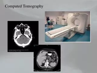
Gantry • Is the movable frame of the CT unit • It contains the x-ray tube and the detectors • It maintain the alignment of the tube and the detectors and contains the equipment necessary to perform the scanning movement • The aperture for the patient is 50-58 cm • Do not force the obese patient into the aperture
Gantry • Most gantrys can be angled up to 30o • Table angulation can be sometimes be used in place of gantry angulation • Positioning lights are usually mounted on the gantry intense white halogen lights and low power laser lights • There are often 3 positioning lights for accurate sagittal, coronal, and transverse centering
X-ray Tubes • CT images produce massive amounts of heat in the x-ray tube • Focal spots size use 0.6 mm and 1.2 mm • 0.6 mm use a pulsed beam to reduce the heat load • Modern CT pulsed scanner tubes operate at 120 kVp, 1-5 msec pulses • X-ray tube produce 0.5-5 million heat unit • Liquid and air-cooled tube housing design have been developed
X-ray Tubes • The radiation beam is double collimated to assist in eliminating scatter information • Slight misalignment can create ring artifact image • The anode is aligned with their long axis perpendicular to the scanner plane, this to prevent heel effect • Collimation controls voxel length • Can be vary between 1mm to 13 mm usually control by the software program • The dimension width determine by section thickness or voxel length
Detectors • CT detectors should have: • (a) high capture efficiency i.e. how well the detectors receive the photon from the patient and that can be control by detectors size and the distance between detectors • (b) high absorption efficiency i.e. how well the detectors convert incoming photons, it determine by the material used as well the size and thickness of the detector
Detectors • (c) high conversion efficiency i.e. how well the detector convert the absorb photon to analog or digital signal • CT detectors should have also high stability, fast response time, and wide dynamic range which is the ratio between the largest signal to the smallest that can be measured • Typical modern scanners are capable of dynamic range of 1,000,000:1
Computer • Is design to control the data acquisition, process and display, and storage • The computer should be in enclosed room with controlled temperature and humidity • CT console provide the radiographer access to the software program that data acquisition, controls data processing and display and storage functions
Computer • A system program is used to start up the CT unit, this program turns on and perform quality assurance, and record various problems • The CT console operate from the menu simply uses a keyboard, light pen
Data Acquisition • Controls the tube and detector collimation (pixel size), matrix size, gantry angle, table top entrance, section increment movement, kVp, mA, scan speed
Display Console • Controls the digital image production process, that compile the image and display parameters, such as window width and level
Exposure Factor • Most CT performed at 120 kVp • Time is not a factor as it must be controlled by scanning program • mA should be setup • Dual energy scanning units required usually 80 kVp and 140 kVp
Artifacts • Motion • Metal or Star • Beam Hardening • Partial Volume Effect • Ring artifacts