Download
1 / 34
360 likes | 436 Vues
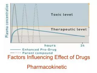
Understand the process of drug distribution in the body, factors influencing it, and the dynamics of plasma to tissue transfer. Learn about cardiac output, capillary permeability, and binding to proteins affecting drug distribution.

E N D
Pharmacokinetic Assistant Professor Dr. Hayder B Sahib Ph.D., M.Sc., D.Sc., B.Sc. Pharm
IV. DRUG DISTRIBUTION • Drug distribution is the process by which a drug reversibly leaves the bloodstream and enters the interstitium (extracellular fluid) and the tissues. • For drugs administered IV, absorption is not a factor, and the initial phase (from immediately after administration through the rapid fall in concentration) represents the distribution phase, during which the drug rapidly leaves the circulation and enters the tissues.
The distribution of a drug from the plasma to the interstitium depends on • 1- Cardiac output • 2- Local blood flow • 3- Capillary permeability • 4- The tissue volume • 5- The degree of binding of the drug to plasma and tissue proteins • 6- The relative lipophilicity of the drug.
A. Blood flow • The rate of blood flow to the tissue capillaries varies widely. For instance, blood flow to the “vessel-rich organs” (brain, liver, and kidney) is greater than that to the skeletal muscles. • Adipose tissue, skin, and viscera have still lower rates of blood flow. • Variation in blood flow partly explains the short duration of hypnosis produced by an IV bolus of propofol. • High blood flow, together with high lipophilicity of propofol, permits rapid distribution into the CNS and produces anesthesia. • A subsequent slower distribution to skeletal muscle and adipose tissue lowers the plasma concentration so that the drug diffuses out of the CNS, down the concentration gradient, and consciousness is regained.
B. Capillary permeability • Capillary permeability is determined by • 1- capillary structure • 2-chemical nature of the drug. • Capillary structure varies in terms of the fraction of the basement membrane exposed by slit junctions between endothelial cells. • In the liver and spleen, a significant portion of the basement membrane is exposed due to large, discontinuous capillaries through which large plasma proteins can pass). • In the brain, the capillary structure is continuous, and there are no slit junctions).
To enter the brain, drugs must pass through the endothelial cells of the CNS capillaries or be actively transported. • For example, a specific transporter carries levodopa into the brain. • By contrast, lipid-soluble drugs readily penetrate the CNS because they dissolve in the endothelial cell membrane. • Ionized or polar drugs generally fail to enter the CNS because they cannot pass through the endothelial cells that have no slit junctions. • These closely juxtaposed cells form tight junctions that constitute the blood–brain barrier.
C. Binding of drugs to plasma proteins and tissues • 1. Binding to plasma proteins: • Reversible binding to plasma proteins isolates drugs in a nondiffusible form and slows their transfer out of the vascular compartment. • Albumin is the major drug-binding protein and may act as a drug reservoir (as the concentration of free drug decreases due to elimination, the bound drug dissociates from the protein). • This maintains the freedrug concentration as a constant fraction of the total drug in the plasma.
Binding to tissue proteins: • Many drugs accumulate in tissues, leading to higher concentrations in tissues than in the extracellular fluid and blood. • Drugs may accumulate as a result of binding to lipids, proteins, or nucleic acids. • Drugs may also be actively transported into tissues. Tissue reservoirs may serve as a major source of the drug and prolong its actions or cause local drug toxicity. (For example, acrolein, the metabolite of cyclophosphamide, can cause hemorrhagic cystitis because it accumulates in the bladder.
D. Lipophilicity • The chemical nature of a drug strongly influences its ability to cross cell membranes. • Lipophilic drugs readily move across most biologic membranes. • These drugs dissolve in the lipid membranes and penetrate the entire cell surface. • The major factor influencing the distribution of lipophilic drugs is blood flow to the area. In contrast, hydrophilic drugs do not readily penetrate cell membranes and must pass through slit junctions.
E. Volume of distribution • The apparent volume of distribution, Vd, is defined as the fluid volume that is required to contain the entire drug in the body at the same concentration measured in the plasma. • It is calculated by dividing the dose that ultimately gets into the systemic circulation by the plasma concentration at time zero (C0).
1. Distribution into the water compartments in the body: • Once a drug enters the body, it has the potential to distribute into any one of the three functionally distinct compartments of body water or to become sequestered in a cellular site. • A. Plasma compartment: If a drug has a high molecular weight or is extensively protein bound, it is too large to pass through the slit junctions of the capillaries and, thus, is effectively trapped within the plasma (vascular) compartment. • As a result, it has a low Vd that approximates the plasma volume or about 4 L in a 70-kg individual. Heparin shows this type of distribution.
B. Extracellular fluid: If a drug has a low molecular weight but is hydrophilic, it can pass through the endothelial slit junctions of the capillaries into the interstitial fluid. • However, hydrophilic drugs cannot move across the lipid membranes of cells to enter the intracellular fluid. • Therefore, these drugs distribute into a volume that is the sum of the plasma volume and the interstitial fluid, which together constitute the extracellular fluid (about 20% of body weight or 14 L in a 70-kg individual). • Aminoglycoside antibiotics show this type of distribution.
C. Total body water: If a drug has a low molecular weight and is lipophilic, it can move into the interstitium through the slit junctions and also pass through the cell membranes into the intracellular fluid. • These drugs distribute into a volume of about 60% of body weight or about 42 L in a 70-kg individual. Ethanol exhibits this apparent Vd
2. Apparent volume of distribution: A drug rarely associates exclusively with only one of the water compartments of the body. • Instead, the vast majority of drugs distribute into several compartments, often avidly binding cellular components, such as lipids (abundant in adipocytes and cell membranes), proteins (abundant in plasma and cells), and nucleic acids (abundant in cell nuclei). • Therefore, the volume into which drugs distribute is called the apparent volume of distribution (Vd). • Vd is a useful pharmacokinetic parameter for calculating the loading dose of a drug.
3. Determination of Vd: The fact that drug clearance is usually a first-order process allows calculation of Vd. • First order means that a constant fraction of the drug is eliminated per unit of time. • This process can be most easily analyzed by plotting the log of the plasma drug concentration (Cp) versus time. • Vd= Dose /PC
4. Effect of Vd on drug half-life: • Vd has an important influence on the half-life of a drug, because drug elimination depends on the amount of drug delivered to the liver or kidney (or other organs where metabolism occurs) per unit of time. • Delivery of drug to the organs of elimination depends not only on blood flow but also on the fraction of the drug in the plasma. • If a drug has a large Vd, most of the drug is in the extra plasmic space and is unavailable to the excretory organs. Therefore, any factor that increases Vd can increase the half-life and extend the duration of action of the drug.
V. DRUG CLEARANCE THROUGH METABOLISM • Once a drug enters the body, the process of elimination begins. • The three major routes of elimination are • 1- hepatic metabolism • 2- biliary elimination • 3- urinary elimination. • Together, these elimination processes decrease the plasma concentration exponentially. • Most drugs are eliminated according to first-order kinetics, although some, such as aspirin in high doses, are eliminated according to zero-order or nonlinear kinetics.
Metabolism leads to production of products with increased polarity, which allows the drug to be eliminated. • Clearance (CL) estimates the amount of drug cleared from the body per unit of time. • Total CL is a composite estimate reflecting all mechanisms of drug elimination and is calculated as follows: • CL= 0.693x Vd/t1/2
A. Kinetics of metabolism • 1. First-order kinetics: The metabolic transformation of drugs is catalyzed by enzymes, and most of the reactions obey Michaelis Menten kinetics. • V=Rate of drug metabolism= Vmax[c] • km+[c] • In most clinical situations, the concentration of the drug, [C], is much less than the Michaelis constant, Km, and the MichaelisMenten equation reduces to
That is, the rate of drug metabolism and elimination is directly proportional to the concentration of free drug, and first-order kinetics is observed. • This means that a constant fraction of drug is metabolized per unit of time (that is, with each half-life, the concentration decreases by 50%). • First-order kinetics is also referred to as linear kinetics.
2. Zero-order kinetics: • With a few drugs, such as aspirin, ethanol, and phenytoin, the doses are very large. Therefore, [C] is much greater than Km • The enzyme is saturated by a high free drug concentration, and the rate of metabolism remains constant over time. • This is called zero-order kinetics (also called nonlinear kinetics). A constant amount of drug is metabolized per unit of time. • The rate of elimination is constant and does not depend on the drug concentration.
B. Reactions of drug metabolism • The kidney cannot efficiently eliminate lipophilic drugs that readily cross cell membranes and are reabsorbed in the distal convoluted tubules. Therefore, lipid-soluble agents are first metabolized into more polar (hydrophilic) substances in the liver via two general sets of reactions, called phase I and phase II
1. Phase I • : Phase I reactions convert lipophilic drugs into more polar molecules by introducing or unmasking a polar functional group, such as –OH or –NH2. • Phase I reactions usually involve reduction, oxidation, or hydrolysis. • Phase I metabolism may increase, decrease, or have no effect on pharmacologic activity.
a. Phase I reactions utilizing the P450 system: The phase I reactions most frequently involved in drug metabolism are catalyzed by the cytochrome P450 system (also called microsomal mixed-function oxidases). • The P450 system is important for the metabolism of many endogenous compounds (such as steroids, lipids) and for the biotransformation of exogenous substances (xenobiotics). • Cytochrome P450, designated as CYP, is a superfamily of heme-containing isozymes that are located in most cells, but primarily in the liver and GI tract.
[1] Nomenclature: The family name is indicated by the Arabic number that follows CYP, and the capital letter designates the subfamily, for example, CYP3A. • A second number indicates the specific isozyme, as in CYP3A4. • [2] Specificity: Because there are many different genes that encode multiple enzymes, there are many different P450 isoforms. These enzymes have the capacity to modify a large number of structurally diverse substrates. In addition, an individual drug may be a substrate for more than one isozyme. • Four isozymes are responsible for the vast majority of P450-catalyzed reactions. They are CYP3A4/5, CYP2D6, CYP2C8/9, and CYP1A2. Considerable amounts of CYP3A4 are found in intestinal mucosa, accounting for first-pass metabolism of drugs such as chlorpromazine and clonazepam.
[3] Genetic variability: P450 enzymes exhibit considerable genetic variability among individuals and racial groups. • Variations in P450 activity may alter drug efficacy and the risk of adverse events. CYP2D6, in particular, has been shown to exhibit genetic polymorphism. CYP2D6 mutations result in very low capacities to metabolize substrates. • Some individuals, for example, obtain no benefit from the opioid Oxidation
analgesic codeine, because they lack the CYP2D6 enzyme that activates the drug. Similar polymorphisms have been characterized for the CYP2C subfamily of isozymes. • For instance, clopidogrel carries a warning that patients who are poor CYP2C19 metabolizers have a higher incidence of cardiovascular events (for example, stroke or myocardial infarction) when taking this drug. • Clopidogrel is a prodrug, and CYP2C19 activity is required to convert it to the active metabolite. • Although CYP3A4 exhibits a greater than 10-fold variability between individuals, no polymorphisms have been identified so far for this P450 isozyme.
[4] Inducers: The CYP450-dependent enzymes are an important target for pharmacokinetic drug interactions. One such interaction is the induction of selected CYP isozymes. • Xenobiotics (chemicals not normally produced or expected to be present in the body, for example, drugs or environmental pollutants) may induce the activity of these enzymes. • Certain drugs (for example, phenobarbital, rifampin, and carbamazepine) are capable of increasing the synthesis of one or more CYP isozymes. • This results in increased biotransformation of drugs and can lead to significant decreases in plasma concentrations of drugs metabolized by these CYP isozymes, with concurrent loss of pharmacologic effect.
For example, rifampin, an antituberculosis drug, significantly decreases the plasma concentrations of human immunodeficiency virus (HIV) protease inhibitors, thereby diminishing their ability to suppress HIV replication. • St. John’s wort is a widely used herbal product and is a potent CYP3A4 inducer. Many drug interactions have been reported with concomitant use of St. John’s wort. • 1) decreased plasma drug concentrations • 2) decreased drug activity if the metabolite is inactive • 3) increased drug activity if the metabolite is active • 4) decreased therapeutic drug effect.
[5] Inhibitors: Inhibition of CYP isozyme activity is an important source of drug interactions that lead to serious adverse events. • The most common form of inhibition is through competition for the same isozyme. Some drugs, however, are capable of inhibiting reactions for which they are not substrates (for example, ketoconazole), leading to drug interactions. • Numerous drugs have been shown to inhibit one or more of the CYP-dependent biotransformation pathways of warfarin. • For example, omeprazole is a potent inhibitor of three of the CYP isozymes responsible for warfarin metabolism. If the two drugs are taken together, plasma concentrations of warfarin increase, which leads to greater anticoagulant effect and increased risk of bleeding.
The more important CYP inhibitors are erythromycin, ketoconazole, and ritonavir, because they each inhibit several CYP isozymes. • Natural substances may also inhibit drug metabolism. For instance, grapefruit juice inhibits CYP3A4 and leads to higher levels and/or greater potential for toxic effects with drugs, such as nifedipine, clarithromycin, and simvastatin, that are metabolized by this system.
b. Phase I reactions not involving the P450 system: These include amine oxidation (for example, oxidation of catecholamines or histamine), alcohol dehydrogenation (for example, ethanol oxidation), esterases (for example, metabolism of aspirin in the liver), and hydrolysis (for example, of procaine). • 2. Phase II: This phase consists of conjugation reactions. If the metabolite from phase I metabolism is sufficiently polar, it can be excreted by the kidneys. • However, many phase I metabolites are still too lipophilic to be excreted. • A subsequent conjugation reaction with an endogenous substrate, such as glucuronic acid, sulfuric acid, acetic acid, or an amino acid, results in polar, usually more water-soluble compounds that are often therapeutically inactive.
A notable exception is morphine-6-glucuronide, which is more potent than morphine. Glucuronidation is the most common and the most important conjugation reaction. [Note: Drugs already possessing an –OH, –NH2, or –COOH group may enter phase II directly and become conjugated without prior phase I metabolism.] • The highly polar drug conjugates are then excreted by the kidney or in bile.