Chapter 19 Oxidative phosphorylation and photophosphorylation
1.2k likes | 1.78k Vues
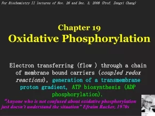
Chapter 19 Oxidative phosphorylation and photophosphorylation. Generation of ATP by using a across-membrane proton gradient, which is generated from electron flowing through a chain of carriers.

Chapter 19 Oxidative phosphorylation and photophosphorylation
E N D
Presentation Transcript
Chapter 19 Oxidative phosphorylation and photophosphorylation Generation of ATP by using a across-membrane proton gradient, which is generated from electron flowing through a chain of carriers.
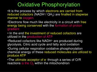
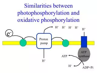
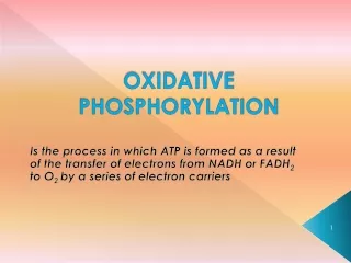
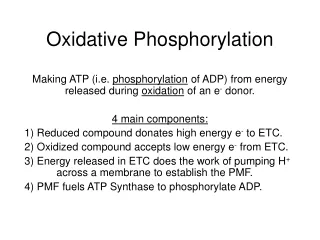
1. ATP is synthesized using the same strategy in oxidative phosphorylation and photophosphorylation • Oxidative phosphorylation is the process in which ATP is generated as a result of electron flow from NADH or FADH2 to O2 via a series of membrane-bound electron carriers, called the respiratory chain (reducing O2 to H2O at the end). • Photophosphorylation is the process in which ATP (and NADPH) is synthesized as a result of electron flow from H2O to NADP+ via a series of membrane-bound electron carriers (oxidizing H2O to O2 at the beginning).
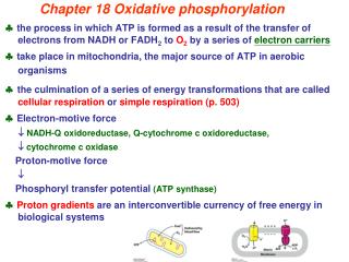
Oxidative phosphorylation and photophosphorylation are mechanistically similar: • Both involve the flow of electrons through a chain of membrane-bound carriers. • The energy released from “downhill” electron flow is first used for “uphill” pumping of protons to produce a proton gradient (thus a transmembrane electrochemical potential) cross a biomembrane. • ATP is then synthesized by a “downhill” transmembrane flow of protons through a specific protein machinery.
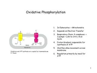
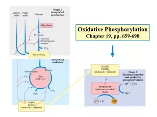
Cristae (the convoluted inner membrane of mitochondria) is where the respiratory chain is located.
Oxidative Phosphorylation (0n inner membrane of mitochondria)
Photophosphorylation (on thylakoid of chloroplasts)
2. Electrons collected in NADH and FADH2 are released and transported to O2via the respiratory chain • The chain is located on the convoluted inner membrane (cristae) of mitochondria in eukaryotic cells (revealed by Eugene Kennedy and Albert Lehninger in 1948) or on the plasma membrane in prokaryotic cells. • A 1.14-volt potential difference (E`0) between NADH (-0.320 V) and O2 (0.816 V) drives electron flow through the chain.
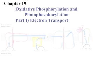
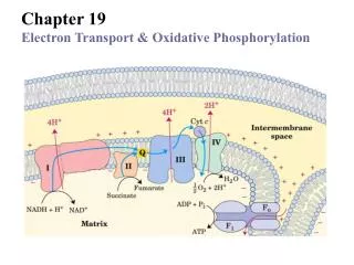
The respiratory chain consists of four large multi-protein complexes (I, II, III, and IV; three being proton pumps) and two mobile electron carriers, ubiquinone (Q or coenzyme Q) and cytochrome c. • Prosthetic groups acting in the proteins of respiratory chain include flavins (FMN, FAD), hemes (heme A, iron protoporphyrin IX, heme C), iron-sulfur clusters (2Fe-2S, 4Fe-4S), and copper.
I II III Four multi-protein Complexes (I, II, III, and IV) Two mobile Electron carriers IV
FMN can accept one electron ( and FMNH2 can donate one electron) to form a semiquinone radical intermediate.
Heme groups of cytochrome proteins Heme groups Of cytochromes
2Fe-2S 4Fe-4S Different types of iron-sulfur centers A ferredoxin • Iron atoms cycle between Fe2+ (reduced) and Fe3+(oxidized).
Reduced cytochromes has three absorption bands in the visible wavelengths Cyt a: ~600 nm; Cyt b: ~560 nm; Cyt c: ~550 nm
3. NADH enters the chain at NADH dehydrogenase (complex I) • Also named as NADH:ubiquinone oxidoreductase or NADH-Q reductase. • A “L” shaped 850 kD multimeric protein complex of 42 different subunits (larger than a ribosome!). • Polypeptides encoded by both genomes. • FMN, Fe-S centers act as prosthetic groups. • Exergonic electron transferring is coupled to endergonic proton pumping ( with 4 H+ pumped from the matrix side to intermembrane space per electron pair transferred), with mechanism unknown. • Final electron acceptor is ubiquinone (coenzyme Q).
NADH Dehydrogenase (complex I)
Ubiquinone is a mobile electron/proton carrier • Fat soluble benzoquinone with a very long isoprenoid side chain; can accept one or two electrons, forming radical semiquinone or ubiquinol (QH2); QH2 diffuses to the next complex (III); the only electron carrier not bound to a protein.
4. FADH2 of flavoproteins also transfer their electrons to ubiquinone • Flavoproteins like succinate dehydrogenase (complex II), fatty acyl-CoA dehydrogenase, and glycerol 3-phosphate dehydrogenase are associated to the inner membrane of mitochondria and transfers their electrons collected on FADH2 to Q to form QH2. • The energy released from these electron transferring is not high enough to promote proton pumping.
Ubiquinone (Q) accepts electrons from both NADH and FADH2 in the respiratory chain
5. Electrons of QH2 is transferred to cytochrome c via ubiquinone:cytochrome c oxidoreductase (complex III) • Also called cytochrome c reductase or cytochrome bc1 complex. • A 250 kD multiprotein complex of 11 subunits. • Complete 3-D structure was determined in 1997! • The functional core consists of three subunits: cytochrome b (with two hemes, bH and bL); an Fe-S protein; and cytochrome c1 (with the heme group covalently bound to protein via two thioether bonds).
Two-electron carrier QH2 passes one electron to the one-electron carrier Fe-S center, then to the heme C group in Cyt c1, and finally to the heme C group of Cyt c; the other electron to bL, bH, and finally to an Q or Q.- via a so-called “Q cycle”. • Cytochrome c, a soluble protein located in the intermembrane space, will move to complex IV.
The three core subunits Cytochrome bc1 complex (complex III)
Electron path in complex III The Q cycle The Q cycle The 1st QH2 The 2nd QH2
6. Electrons of Cyt c are transferred to O2 on cytochrome oxidase (complex IV) • A 204 kD 13-subunit protein complex, with structure determined in 1996. • Three subunits are probably critical to the function. • Three copper ions (2 CuA, 1CuB), two heme A groups (a and a3) act as electron carriers in complex IV. • Four electrons need to be transferred to reduce one O2 molecule at the “Fe-CuB center” (via peroxy intermediates) of complex IV to form 2 H2O.
Four “substrate” protons are consumed from the N side for every four electrons transferred to one O2 molecule. • One proton is pumped out from the N to P side for each electron to be transferred by an yet defined mechanism.
2CuA CuB Heme a CuA Heme a3 CuA The three critical subunits of cytochrome oxidase (complex IV)
The electron path in complex IV
7. A proton gradient across the inner membrane of mitochondria is generated using the electron motive force • An estimate of 10 protons are pumped for oxidizing one NADH and 6 for one FADH2 accompanying the electron flow through complexes I, III and IV. • Conformational changes induced by electron transferring is believed to be coupled to proton pumping (however, the actual mechanisms is yet revealed!). • In actively respiring mitochondria, the measured pH is about 0.75 and difference in electrical potential () is about 0.15-0.2 V.
The energy stored in such an H+ gradient can be used to synthesize ATP or to do other work.
A H+ gradient across the inner membrane of mitochondria (or plasma membrane of bacteria) is generated by “uphill” H+ pumping using energy released by the “downhill” flow of electrons.
8. The order of the many electron carriers on the respiratory chain have been elucidated via various studies • Measurement of the standard reduction potential (E`0)): Electrons tend to transfer from low E`0 carriers to high E`0 carriers (but may deviate from this in real cells). • Oxidation kinetics studies: Full reduction followed by sudden O2 introduction; earlier oxidation, closer to the end of the respiratory chain; using rapid and sensitive spectrophotometric techniques to follow the oxidation of the cytochromes, which have different wavelength of maximal absorption).
Effects of various specific inhibitors: those before the blocked step should be reduced and those after be oxidized. • Isolation and characterization of each of the multiprotein complexes: specific electron donors and acceptors can be determined for portions of the chain.
Oxidized Reduced Reduced Oxidized Reduced Various inhibitors generate various patterns of reduced/oxidized carriers
9. Electron transfer to O2 was found to be coupled to ATP synthesis from ADP + Pi in isolated mitochondria • ATP would not be synthesized when only ADP and Pi are added in isolated mitochondria suspensions. • O2 consumption, an indication of electron flow, was detected when a reductant (e.g., succinate) is added, accompanied by an increase of ATP synthesis. • Both O2 consumption and ATP synthesis were suppressed when inhibitors of respiratory chain (e.g., cyanide, CO, or antimycin A) was added. • ATP synthesis depends on the occurrence of electron flow in mitochondria.
O2 consumption was neither observed if ADP was not added to the suspension, although a reductant is provided. • The O2 consumption was also not observed in the presence of inhibitors of ATP synthase (e.g., oligomycin or venturicidin). • Electron flow also depends on ATP synthesis!
Electron transfer was found to be obligatorily coupled to ATP Synthesis in isolated mitochondria suspensions: neither occurs without the other.
10. It was widely believed that ATP synthesis occurs by chemical coupling • High energy intermediates similar to 1,3-bisphophoglycerate (which is formed in the glycolytic pathway and transfers an phosphoryl group to ADP to form ATP) was proposed to be produced first from the electron flows on both the mitochondrial and chloroplast membranes. • Phophorylated protein intermediates were also hypothesized. • But neither were ever revealed despite intense efforts by a large number of investigators over many years.
11. The chemiosmotic model was proposed to explain the coupling of electron flow and ATP synthesis • First proposed in 1961 by Peter Michell (a British). • Energy released from electron transferring is hypothesized to be first used to pump protons from the mitochondrial matrix to the intermembrane space (or from stroma to thylakoid lumen in chloroplasts), thus generating a proton gradient across the inner membrane; such a proton-motive force then drives ATP synthesis by moving protons back into the matrix via the ATP synthase.
The model was initially opposed by virtually all researchers working in oxidative phosphorylation and photosynthesis.
The chemiosmotic model by Mitchell
12. The supporting evidences for the chemiosmotic coupling was collected • A closed membrane system is essential for ATP synthesis but not for the electron flow (tested with detergent or physical shearing. • Hydrophobic weak acids (DNP and FCCP) and ionophores (valinomycin) were found to be able to uncouple ATP synthesis from electron transferring. • The transmembrane proton pumping has been experimentally detected: pH in the intermembrane space was found to decrease when electron flow occurs (more protons are pumped when NADH, rather than succinate, is utilized as reductant).
An artificially imposed electrochemical gradient across the chloroplast thylakoid membrane and inner mitochondrial membrane alone (both were performed using sub-organelle vesicles) were found to drive ATP synthesis (with the ATP synthase present). • The across-membrane proton gradient was thus finally accepted as the driving force for ATP synthesis: the chemiosmotic model was accepted as a theory! • The chemiosmotic theory unified the apparently disparate energy transduction processes as oxidative phosphorylation, photophosphorylation, active transport across membrane and the motion of bacterial flagella.
ATP synthesized DNP, a hydrophobic weak acid, uncouples ATP synthesis from electron flow
DNP and CCCP are able to dissipate the proton gradient
The artificially imposed proton gradient alone was found to drive ATP synthesis!
13. ATP synthase was first identified by dissociation and reconstitution studies • Abundant knoblike protruding structures were observed on the matrix side of the inner mitochondrial membrane by EM (Racker in 1960). • The inside-out submitochondrial particles with the “knobs” are capable of both electron transferring and ATP synthesis. • When the protruding F1 part was removed by agitation, electron transferring could still occur, but without proton gradient and ATP synthesis.