Download
1 / 27
320 likes | 1.22k Vues
Chapter 19 Oxidative Phosphorylation and Photophosphorylation. Oxidative Phosphorylation In mitochondria Reduction of O 2 to H 2 O with electrons from NADH or FADH 2 Independent on the light energy Photophosphorylation In chloroplast

E N D
Chapter 19 Oxidative Phosphorylation and Photophosphorylation
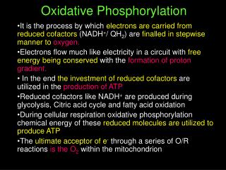
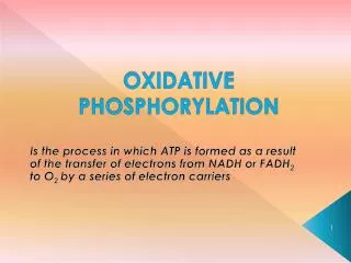
Oxidative Phosphorylation • In mitochondria • Reduction of O2 to H2O with electrons from NADH or FADH2 • Independent on the light energy Photophosphorylation • In chloroplast • Oxidation of H2O to O2 with NADP+ as electron acceptor • Dependent on the light energy
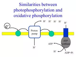
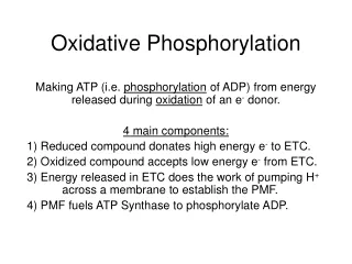
Oxidative Phosphorylation vs. Photophosphorylation • Similarities • Flow of electrons through a chain of membrane-bound carriers (Downhill: exogernic process) • Proton transport across a proton-impermeable membrane (Uphill: endogernic process) Free energy from electron flow is coupled to generation of proton gradient across membrane • Transmembrane electrochemical potential (conserving free energy of fuel oxidation) “Chemiosmotic theory by Peter Mitchell (1961)” • Proton gradient as a reservoir of energy generated by biological oxidation • ATP synthase couples proton flow to ATP synthesis
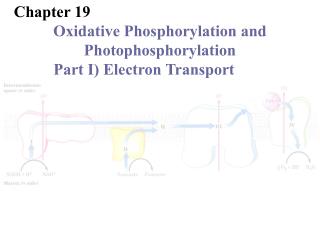
Oxidative Phosphorylation 19.1 Electron-Transfer Reactions in Mitochondria
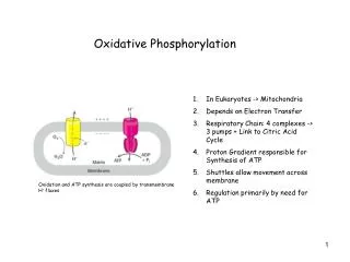
Mitochondria • Site of oxidative phosphorylation • Eugene Kennedy and Albert Lehninger (1948) • Structure • Outer membrane • Free diffusion of small molecules (Mr < 5,000) and ions through porin channels • Inner membrane • Impermeable to most small molecules and ions (protons) • Selective transport • Components of the respiratory chain and the ATP synthase • Mitochondria matrix • Contain enzymes for metabolism • Pyruvate dehydrogenase complex • Citric acid cycle • b-oxidation • Amino acid oxidation
Electron transfer in biological system • Types of electron transfer in biological system • Direct electron transfer : Fe3+ Fe2+ • Hydrogen atom (H+ + e-) • Hydride ion (:H-) • Organic reductants * Reducing equivalent • A single electron equivalent transferred in an redox reaction • Types of electron carriers • NAD(P)+ • FAD or FMN • Ubiquinone (coenzyme Q , Q) • Cytochrome • Iron-sulfur proteins
NAD(P)+ & FAD/FMN ; universal electron acceptors • NAD(P)+ • Cofactors of dehydrogenases (generally) • Electron transfer as a form of :H- • Low [NADH]/[NAD+] catabolic reactions • High [NADPH]/[NADP+] anabolic reactions • No transfer into mito matrix • Shuttle systems (inner mito membrane) Full reduction; 360nm absorption Partial reduction; 450nm absorption • FAD/FMN (flavin nucleotides) • Tightly bound in flavoprotein (generally) • One (semiquinone) or two (FADH2 or FMNH2) electron accept • High reduction potential (induced by binding to protein) Full oxidation; 370 & 440 nm absorption
Membrane-bound electron carriers ; Ubiquinone • Coenzyme Q or Q • Lipid-soluble benzoquinone with long isoprenoid side chain • Accept one (semiquinone radical; •QH) or two electrons (ubiquinol; QH2) • Freely diffusible within inner mito membrane • Shuttling reducing equivalents between less mobile electron carriers • Coupling electron flow to proton movement
Membrane-bound electron carriers ; Cytochromes • Iron-containing heme prosthetic group • 3 classes of Cyt in mitochondria (depending on differences in light-absorption spectra) ; a (near 600nm), b (near 560nm), c (near 550nm) • Cyt c - Covalently-attached heme through Cys - Soluble protein associated with outer surface of inner mito membrane
Membrane-bound electron carriers ; Iron-sulfur proteins • Irons associated with inorganic S or S of Cys • One electron transfer by redox reaction of one iron atom • > 8 Fe-S proteins involved in mito electron transfer • Reduction potential of the protein : -0.65 V ~ +0.45 V
Determining the Sequence of Electron Transfer Chain • Based on the order of standard reduction potential (E’°) • Electron flow from lower E’°tohigher E’° • NADH Q Cyt b Cyt c1 Cyt c Cyt a Cyt a3 O2
Determining the Sequence of Electron Transfer Chain • Reduction of the entire chain of carriers sudden addition of O2 • Spectroscopic measurement of oxidation of each electron carriers • Closer to O2 faster oxidation • Inhibitors • Blocking the flow of electrons • Before/after the inhibited step : fully reducted/ fully oxdized
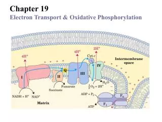
Electron Carriers in multienzyme complex • Membrane-embedded supramolecular complexes (organized in mito respiratory chain) • Complex I : NADH Q • Complex II : Succinate Q • Complex III : Q Cyt c • Complex IV : Cyt to O2 • Separation of functional complexes of respiratory chain
Complex I : NADH:ubiquinone oxidoreductase (NADH dehydrogenase) • 42 polypeptide chains • FMN-containing flavoprotein • > 6 iron sulfur centers • Functions : proton pump driven by the energy from electron transfer • Exergonic transfer of :H- from NADH and a proton from the matrix to Q • NADH + H+ + Q NAD+ + QH2 • Endergonic transfer 4 H+ from the matrix to the intermembrane space • NADH + 5HN+ + Q NAD+ + QH2 + 4Hp+ • Inhibitors : e- flow from Fe-S center • Amytal (a barbiturate drug) • Rotenone (plant, insecticide) • Piericidin A (antibiotic)
Complex II : Succinate Dehydrogenase • Only membrane-bound enzyme in the citric acid cycle • Structure • 4 subunits • C and D : transmembrane side • Heme b : preventing electron leakage to form reactive oxygen species • Q binding site • A and B : matrix side • Three 2Fe-2S centers • FAD • Binding site of succinate • Electron passage : entirely 40 Å long (< 11 Å of each step)
Electron transfer from Glycerol 3-phosphate & fatty acyl-CoA • Electron from fatty acyl-CoA • FAD electron-transferring flavoprotein (ETF) ETF: ubiquinone oxidoreductase Q • Electron from glycerol 3-phosphate • FAD in glycerol 3-phosphate dehydrogenase Q
Shuttling reducing equivalents from cytosolic NADH into mito matrix • ; glycerol 3-phosphate dehydrogenase
Complex III: Cyt bc1 complex (Q:Cyt c oxidoreductase) • e- transfer (ubiquinol (QH2) Cyt c) H+ transfer (matrix intermembrane space) • Dimer of identical monomers (each with 11 different subunits) • Functional core of each monomer; cyt b (2 heme; bH & bL) + Rieske iron-sulfur protein (2Fe-2S center) + cyt c1 (heme c1)
Complex III: Cyt bc1 complex (Q:Cyt c oxidoreductase) • Two binding sites for ubiquinone • ; QN & QP • Antimycin A: binding at QN block e- flow (heme bH Q) • Myothiazol: binding at QP block e- flow (QH2 Rieske iron-sulfur protein) • Cavern (space at the interface between monomers) • ; QN & QP are located
Q cycle in complex III • Two stages • 1st stage; Q (on N side) semiquinone radical • 2nd stage; semiquinone radical QH2
Complex IV : Cytochrome Oxidase • e- transfer from cyt c to O2 H2O • Structure; 13 subunits • Subunit II; 2 Cu ions complexed with –SH of 2 Cys (CuA) 1st binuclear center • Subunit I; 2 heme groups, a & a3 Cu ion (CuB) a3 + CuB 2nd binuclear center
Complex IV : Cytochrome Oxidase • Electron transfer • Cyt c CuA heme a heme a3-CuB center O2 • 4 Cyt c (red) + 8 HN+ + O2 4 cyt c (ox) + 4Hp+ + 2 H2O • 4HN+ as substrate, 4HN+ forpumping out