Insights into Oxidative Phosphorylation: The Powerhouse of ATP Generation
Learn about oxidative phosphorylation, the process by which ATP is generated from the transfer of electrons in mitochondria. Discover the biochemistry behind electron-transfer reactions, electron carriers, and the intricate pathways involved. Delve into the functions of cytochromes, iron-sulfur proteins, and mitochondrial complexes in energy transductions.

Insights into Oxidative Phosphorylation: The Powerhouse of ATP Generation
E N D
Presentation Transcript
Oxidative Phosphorylation Dr. Fayez Almabhouh Assistant Professor, Biology and Biotechnology Department
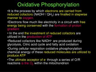
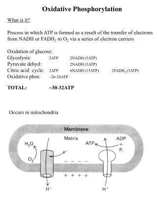
Oxidative phosphorylation • The NADH and FADH2 formed in glycolysis, fatty acid oxidation, and the citric acid cycle are energy-rich molecules because each contains a pair of electrons having a high transfer potential. • When these electrons are used to reduce molecular oxygen to water, a large amount of free energy is liberated, which can be used to generate ATP.
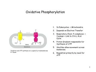
Oxidative phosphorylation • Oxidative phosphorylation is the process in which ATP is formed as a result of the transfer of electrons from NADH or FADH2 to O2by a series of electron carriers. • This process, which takes place in mitochondria, is the major source of ATPin aerobic organism
Electron-Transfer Reactions in Mitochondria • The discovery in 1948 by Eugene Kennedy and Albert Lehninger that mitochondria are the site of oxidative phosphorylation in eukaryotes marked the beginning of the modern phase of studies in biological energy transductions.
Electrons Are Funneled to Universal Electron Acceptors • Oxidative phosphorylation begins with the entry of electrons into the respiratory chain. • Most of these electrons arise from the action of dehydrogenases that collect electrons from catabolic pathways and funnel them into universal electron acceptors—nicotinamide nucleotides (NAD+ or NADP+) or flavin nucleotides (FMN or FAD).
Electrons Pass through a Series of Membrane-Bound Carriers • The mitochondrial respiratory chain consists of a series of sequentially acting electron carriers, most of which are integral proteins with prosthetic groups capable of accepting and donating either one or two electrons.
Three types of electron transfers occur in oxidative phosphorylation: • Direct transfer of electrons, as in the reduction of Fe3+ to Fe2+. • Transfer as a hydrogen atom (H+ + e-); • Transfer as a hydride ion (:H-), which bears two electrons.
In addition to NAD and flavoproteins, three other types of electron-carrying molecules function in the respiratory chain: • A hydrophobicquinone (ubiquinone) and • Two different types of iron-containing proteins (cytochromes and iron-sulfur proteins).
Ubiquinone Also called coenzyme Q, or simply Q is a lipid-soluble benzoquinone with a long isoprenoid side chain
Ubiquinonecan accept one electron to become the semiquinone radical (.QH) or two electrons to form ubiquinol (QH2). • Because ubiquinone is both small and hydrophobic, it is freely diffusible within the lipid bilayer of the inner mitochondrial membrane
The cytochromes • The cytochromes are proteins with characteristic strong absorption of visible light, due to their iron containing heme prosthetic groups. • Mitochondria contain three classes of cytochromes, designated a, b, and c, which are distinguished by differences in their light-absorption spectra.
The heme cofactors of a and b cytochromes are tightly, but not covalently, bound to their associated proteins; the hemes of c-type cytochromes are covalently attached through Cys residues
Iron-sulfur proteins • The iron is present not in heme but in association with inorganic sulfur atoms or with the sulfur atoms of Cys residues in the protein, or both. • These iron-sulfur (Fe-S) centers range from simple structures with a single Fe atom coordinated to four Cys-SH groups to more complex Fe-S centers with two or four Fe atoms
Iron-sulfur centers. The Fe-S centers of iron-sulfur proteins may be as simple as (a), with a single Fe ion surrounded by the S atoms of four Cys residues. Other centers include both inorganic and Cys S atoms, as in (b) 2Fe-2S or (c) 4Fe-4S centers
All iron-sulfur proteins participate in one-electron transfers in which one iron atom of the iron-sulfur cluster is oxidized or reduced. • At least eight Fe-S proteins function in mitochondrial electron transfer.
In the overall reaction catalyzed by the mitochondrial respiratory chain, electrons move from NADH, succinate, or some other primary electron donor through flavoproteins, ubiquinone, iron-sulfur proteins, and cytochromes, and finally to O2.
Electron Carriers Function in Multienzyme Complexes • The electron carriers of the respiratory chain are organized into membrane-embedded supramolecular complexes that can be physically separated. • Four electron carrier complexes, each capable of catalyzing electron transfer through a portion of the chain.
Complexes Iand IIcatalyze electron transfer to ubiquinonefrom two different electron donors: NADH (Complex I) and succinate (Complex II). • Complex III carries electrons from reduced ubiquinone to cytochrome c, and Complex IVcompletes the sequence by transferring electrons from cytochrome c to O2.
Complex I: NADH to Ubiquinone • Complex I, also called NADH:ubiquinone oxidoreductase or NADH dehydrogenase, is a large enzyme composed of 42 different polypeptide chains, including an FMN-containing flavoprotein and at least six iron-sulfur centers.
Complex Icatalyzes the transfer of a hydride ion from NADH to FMN, from which two electrons pass through a series of Fe-S centers to the ironsulfur protein N-2 in the matrix arm of the complex.
Electron transfer from N-2 to ubiquinone on the membrane arm forms QH2, which diffuses into the lipid bilayer. • This electron transfer also drives the expulsion from the matrix of four protons per pair of electrons
Complex II: Succinate to Ubiquinone • Complex II is the succinate dehydrogenase, the only membrane-bound enzyme in the citric acid cycle. • Although smaller and simpler than Complex I, it contains five prosthetic groups of two types and four different protein subunits.
Other substrates for mitochondrial dehydrogenases pass electrons into the respiratory chain at the level of ubiquinone, but not through Complex II.
The first step in theβ-oxidation of fatty acyl–CoA, catalyzed by the flavoprotein acyl-CoA dehydrogenase involves transfer of electrons from the substrate to the FAD of the dehydrogenase, then to electron-transferring flavoprotein (ETF), which in turn passes its electrons to ETF: ubiquinone oxidoreductase. This enzyme transfers electrons into the respiratory chain by reducing ubiquinone.
Glycerol 3-phosphate, formed either from glycerol released by triacylglycerol breakdown or by the reduction of dihydroxyacetone phosphate from glycolysis, is oxidized by glycerol 3-phosphate dehydrogenase
This enzyme is a flavoprotein located on the outer face of the inner mitochondrial membrane, and like succinate dehydrogenase and acyl-CoA dehydrogenase it channels electrons into the respiratory chain by reducing ubiquinone
Complex III: Ubiquinone to Cytochrome c • Also called cytochrome bc1 complex or ubiquinone:cytochrome c oxidoreductase, couples the transfer of electrons from ubiquinol (QH2) to cytochrome c with the vectorial transport of protons from the matrix to the intermembrane space.
Cytochrome bc1 complex (Complex III). The complex is a dimer of identical monomers, each with 11 different subunits. • Structure of a monomer. • The functional core is three subunits: cytochrome b (green) with its two hemes (bH and bL, light red); the Rieske iron-sulfur protein (purple) with its 2Fe-2S centers ; and cytochrome c1 (blue) with its heme (red).
The Q cycle. The path of electrons through Complex III is shown by blue arrows. On the P side of the membrane, two molecules of QH2 are oxidized to Q near the P side, releasing two protons per Q (four protons in all) into the intermembrane space. Each QH2 donates one electron (via the Rieske Fe-S center) to cytochrome c1, and one electron (via cytochrome b) to a molecule of Q near the N side, reducing it in two steps to QH2. This reduction also uses two protons per Q, which are taken up from the matrix.
Complex IV: Cytochrome c to O2 • In the final step of the respiratory chain, Complex IV, also called cytochrome oxidase,carries electrons from cytochrome c to molecular oxygen, reducing it to H2O. • Complex IV is a large enzyme (13 subunits) of the inner mitochondrial membrane.
Electron transfer through Complex IV is from • Cytochrome c • CuA center • heme a • hemea3–CuB center • finally to O2 • The three proteins critical to electron flow are subunits I, II, and III. • The larger structure includes the other ten proteins in the complex.
For every four electrons passing through this complex, the enzyme consumes four “substrate” H+ from the matrix (N side) in converting O2 to 2H2O. • It also uses the energy of this redox reaction to pump one proton outward into the intermembrane space (P side) for each electron that passes through. The overall reaction catalyzed by Complex IV is
This four-electron reduction of O2 involves redox centers that carry only one electron at a time, and it must occur without the release of incompletely reduced intermediates such as hydrogen peroxide or hydroxyl free radicals—very reactive species that would damage cellular components. • The intermediates remain tightly bound to the complex until completely converted to water
Summary of the flow of electrons and protons through the four complexes of the respiratory chain. Electrons reach Q through Complexes I and II. QH2 serves as a mobile carrier of electrons and protons. It passes electrons to Complex III, which passes them to another mobile connecting link, cytochrome c. Complex IV then transfers electrons from reduced cytochrome c to O2. Electron flow through Complexes I, III, and IV is accompanied by proton flow from the matrix to the intermembrane space. Recall that electrons from oxidation of fatty acids can also enter the respiratory chain through Q
The Energy of Electron Transfer Is Efficiently Conserved in a Proton Gradient • The transfer of two electrons from NADH through the respiratory chain to molecular oxygen can be written as • NADH + H+ +1/2O2NAD+ + H2O • This net reaction is highly exergonic • A similar for the oxidation of succinate shows that electron transfer from succinate to O2, standard free-energy change
Much of this energy is used to pump protons out of the matrix. • For each pair of electrons transferred to O2, fourprotons are pumped out by Complex I, fourby Complex III, and two by Complex IV. • The vectorial equation for the process is therefore: • NADH + 11HN+ +1/2O2 NAD+ +10HP+ +H2O
The electrochemical energy inherent in this difference in proton concentration and separation of charge represents a temporary conservation of much of the energy of electron transfer. • The energy stored in such a gradient, termed the proton-motive force, has two components: • The chemical potential energy due to the difference in concentration of a chemical species (H+) in the two regions separated by the membrane, and • The electrical potential energy that results from the separation of charge when a proton moves across the membrane
When protonsflow spontaneously down their electrochemical gradient, energy is made available to do work. • In mitochondria, chloroplasts, and aerobic bacteria, the electrochemical energy in the proton gradient drives the synthesis of ATP from ADP and Pi.
ATP Synthesis • How is a concentration gradient of protons transformed into ATP? • Electron transfer releases, and the proton-motive force conserves, more than enough free energy (about 200 kJ) per “mole” of electron pairs to drive the formation of a mole of ATP, which requires about 50 kJ.
What is the chemical mechanism that couples proton flux with phosphorylation?
The chemiosmotic model, proposed by Peter Mitchell, is the paradigm for this mechanism. • According to the model, the electrochemical energy inherent in the difference in proton concentration and separation of charge across the inner mitochondrial membrane—the proton-motive force—drives the synthesis of ATP as protons flow passively back into the matrix through a proton pore associated with ATP synthase.
Chemiosmotic model: The inner mitochondrial membrane is impermeable to protons; protons can reenter the matrix only through proton-specific channels (Fo). The proton-motive force that drives protons back into the matrix provides the energy for ATP synthesis, catalyzed by the F1 complex associated with Fo.
Mitchell used “chemiosmotic” to describe enzymatic reactions that involve, simultaneously, a chemical reaction and a transport process. The operational definition of “coupling” is shown in figure:
Coupling of electron transfer and ATP synthesis in mitochondria. • In experiments to demonstrate coupling, isolatedmitochondria are suspended in a buffered medium containing ADP, Pi, and an oxidizable substrate such as succinate, three easily measured processes occur: (1) The substrate is oxidized (succinate yields fumarate) (2) O2 is consumed, (O2 electrode monitors O2 consumption) (3) ATP is synthesized. • Oxygen consumption and ATP synthesis depend on the presence of an oxidizable substrate (succinate in this case) as well as ADP and Pi.