OXIDATIVE PHOSPHORYLATION
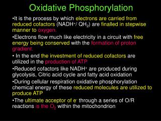
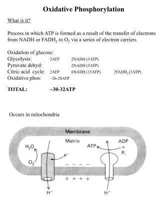
OXIDATIVE PHOSPHORYLATION. Is the process in which ATP is formed as a result of the transfer of electrons from NADH or FADH 2 to O 2 by a series of electron carriers. Takes place in mitochondria Is the major source of ATP in aerobic organisms


OXIDATIVE PHOSPHORYLATION
E N D
Presentation Transcript
OXIDATIVE PHOSPHORYLATION Is the process in which ATP is formed as a result of the transfer of electrons from NADH or FADH2 to O2 by a series of electron carriers
Takes place in mitochondria • Is the major source of ATP in aerobic organisms • Oxidative phosphorylation generates 26 of the 30 molecules of ATP that are formed when glucose is completely oxidized to CO2 and H2O. Mitochondria, Stained Green, Form a Network Inside a Fibroblast Cell
Cellular respiration • Carbon fuels are oxidized in the citric acid cycle to yield electrons with high transfer potential. • This electron-motive force is converted into a proton-motive force. • The proton-motive force is converted into phosphoryl transfer potential.
Mitochondria oxidize carbon fuels to form cellular energy. • This transformation requires electron transfer through several large protein complexes some of which pump protons, forming a proton gradient that powers the synthesis of ATP.
conversion of electron-motive force into proton-motive force • Carried out by three electron-driven proton pumps: • NADH-Q oxidoreductase • Q-cytochrome c oxidoreductase • cytochrome c oxidase. • These large transmembrane complexes contain multiple oxidation-reduction centers, including: • Quinones (Q) • Flavins • Iron-sulfur clusters • Hemes • Copper ion • The final phase of oxidative phosphorylation is carried out by ATP synthase, an ATP-synthesizing assembly driven by the flow of protons back into the mitochondrial matrix.
Essence of Oxidative Phosphorylation • Oxidation and ATP synthesis are coupled by transmembrane proton fluxes.
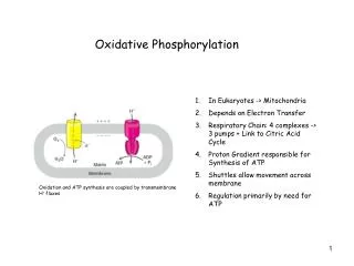
Mitochondria TWO COMPARTMENTS • The intermembrane space between the outer and the inner membranes • Oxidative phosphorylation • The matrix which is bounded by the inner membrane: • most of the reactions of the citric acid cycle and fatty acid oxidation
OUTER MEMBRANE • Permeable to most small molecules and ions because it contains many copies of mitochondrial porin: • VDAC is voltage-dependent anion channel. • VDAC plays a role in the regulated flux of metabolites across the outer membrane : • Phosphate • Chloride • organic anions • adenine nucleotides
VDAC appears to form an open b-barrel structure similar to bacterial porins. • Some cytoplasmic kinases bind to VDAC, thereby obtaining preferential access to the exported ATP.
INNER MEMBRANE • Impermeable to nearly all ions and polar molecules. • A large family of transporters shuttles metabolites across the inner mitochondrial membrane like: • ATP • Pyruvate • citrate • The two faces of this membrane will be referred to as: • matrix side also called the N side (the membrane potential is negative) • cytosolic side (the latter because it is freely accessible to most small molecules in the cytosol). also called the P side (the membrane potential is positive)
High energy electrons: Redox potential & Free-energy change • In oxidative phosphorylation, the electron transfer potential of NADH or FADH2 is converted into the phosphoryl transfer potential of ATP. • Recall that: • DG0’ of hydrolysis of ATP to ADP + Pi gives idea about the phosphoryl transfer potential. • E’0is the electron transfer potential: also called the reduction potential, redox potential or oxidation-reduction potential
A strong reducing agent (such as NADH) is poised to donate electrons and has a negative reduction potential (-ve E’0). • A strong oxidizing agent (such as O2) is ready to accept electrons and has a positive reduction potential (+ve E’0)..
The driving force of oxidative phosphorylation: • Is the electron transfer potential of NADH or FADH2 relative to that of O2. • How much energy is released by the reduction of O2 with NADH? • Subtracting reaction b from reaction a yields: • The standard free energy for this reaction is then given by
Mitochondrial Electron-Transport Chain • Large multi subunit integral membrane protein complexes or coupling sites. • Complex I:NADH-Q oxidoreductase(MW =880 kd) Coupling Site 1 • Complex II:succinate-Q reductase complex (MW =140 kd) • Complex III:Q-cytochrome c oxido-reductase(MW = 250 kd). Coupling Site 2 • Complex IV:cytochrome c oxidase(MW = 160 kd) Coupling Site 3.
Mitochondrial Electron-Transport Chain • Small Mobile Electron Carriers: • Ubiquinone/Ubiquinol (Q/QH2): small hydrophobic electron carriers which shuttle electrons between the large complexes and back and forth across the lipid bilayer. • Cytochrome c: is a small water soluble protein which is a mobile electron carrier and carries electrons between cytochrome bc1 complex and cytochrome c oxidase.
Electron flow within these transmembrane complexes leads to the transport of protons across the inner mitochondrial membrane. • Electrons are carried from NADH-Q oxidoreductase to Q-cytochrome c oxidoreductase by the reduced form of coenzyme Q (Q). • Q also carries electrons from FADH2, generated in succinate dehydrogenase in the citric acid cycle, to Q-cytochrome c oxidoreductase, generated through succinate-Q reductase.
Cytochrome c, a small, soluble protein, shuttles electrons from Q-cytochrome c oxidoreductase to cytochrome c oxidase (complex IV), which catalyzes the reduction of O2. • Succinate-Q reductase (Complex II), in contrast with the other complexes, does not pump protons.
Coenzyme Q (Q) • Q is a hydrophobic quinone that diffuses rapidly within the inner mitochondrial membrane. • Q is a quinone derivative with a long isoprenoid tail. • The number of five-carbon isoprene units in coenzyme Q depends on the species. • The most common form in mammals contains 10 isoprene units (coenzyme Q10).
Oxidation States of Quinones The reduction of ubiquinone (Q) to ubiquinol (QH2) proceeds through a semiquinone anion intermediate (Q.-). The addition of one electron and one proton results in the semiquinone form (QH·). In the fully oxidized state (Q), coenzyme Q has two keto groups. The semiquinone form is relatively easily deprotonated to form a semiquinone radical anion (Q·-). The addition of a second electron and proton generates ubiquinol (QH2),
Thus, electron-transfer reactions of quinones are coupled to proton binding and release, a property that is key to transmembrane proton transport.
Flavins • Oxidation States of Flavins: • The reduction of flavin mononucleotide (FMN) to FMNH2 proceeds through a semiquinone intermediate.
Fe-S clusters • Fe-Sclusters in iron-sulfur proteins (nonheme iron proteins) play a critical role in a wide range of reduction reactions in biological systems. • Several types of Fe-S clusters are known:
A single iron ion bound by four cysteine residues. • 2Fe-2S cluster with iron ions bridged by sulfide ions. • 4Fe-4S cluster. • Each of these clusters can undergo oxidation-reduction reactions.
Iron ions in these Fe-S complexes cycle between: • Fe2+ (reduced state) • Fe3+ (oxidized state) • Unlike quinones and flavins, iron-sulfur clusters generally undergo oxidation-reduction reactions without releasing or binding protons.
COMPLEX INADH-Q oxidoreductase • NADH-Q oxidoreductase (also called NADH dehydrogenase): • an enormous enzyme (880 kd) • consists of at least 34 polypeptide chains. • consists of a membrane-spanning part and a long arm that extends into the matrix. • Contains FMN and Fe-S prosthetic groups.
NADH is oxidized in the arm, and the electrons are transferred to reduce Q in the membrane. • The reaction catalyzed by this enzyme appears to be:
The initial step is the binding of NADH and the transfer of its two high-potential electrons to the flavin mononucleotide (FMN) prosthetic group of this complex to give the reduced form, FMNH2.
Electrons are then transferred from FMNH2 to a series of 4Fe4S, the second type of prosthetic group in NADH-Q oxidoreductase. • NADH-Q oxidoreductase contains: • 2Fe-2S cluster • 4Fe-4S cluster
Electrons in the 4Fe4S of NADH-Q oxidoreductase are shuttled to coenzyme Q. • The flow of two electrons from NADH to coenzyme Q through NADH-Q oxidoreductase leads to the pumping of four hydrogen ions out of the matrix of the mitochondrion. • The coupled electron- proton transfer reactions of Q are crucial.
3 2
3 2
In summary: • NADH binds to a site on the vertical arm and transfers its electrons to FMN. • These electrons flow within the vertical unit to three 4Fe-4S centers. • Then they flow to a bound Q. The reduction of Q to QH2results in the uptake of two protons from the matrix.
The pair of electrons on bound QH2 are transferred to a 4Fe-4S center and the protons are released on the cytosolic side. • These electrons are transferred to a mobile Q in the hydrophobic core of the membrane, resulting in the uptake of two additional protons from the matrix.
COMPLEX II succinate-Q reductase complex • It is an integral membrane protein of the inner mitochondrial membrane. • Contains three different kinds of Fe-S clusters: • 2Fe-2S • 3Fe-4S • 4Fe-4S.
FADH2 is generated in the citric acid cycle by the enzyme succinate dehydrogenase, with the oxidation of succinate to fumarate.
Two electrons are transferred from FADH2 directly to Fe-S clusters of succinate dehydrogenase. • The electrons are then passed to (Q) for entry into the electron-transport chain. • FADH2 is generated by other reactions (like by: Glycerol phosphate dehydrogenase and fatty acyl CoA dehydrogenase). • This FADH2 also transfers its electrons to (Q), to form (QH2).
The succinate-Q reductase complex and other enzymes that transfer electrons from FADH2 to Q, in contrast with NADH-Q oxidoreductase, do not transport protons. • Consequently, less ATP is formed from the oxidation of FADH2 than from NADH.
COMPLEX IIIQ-Cytochrome c Oxidoreductase • The second of the three proton pumps in the respiratory chain (also known as cytochrome reductase). • The function of Q-cytochrome c oxidoreductase is to catalyze the transfer of electrons from QH2 to oxidized cytochrome c (cyt c), a water-soluble protein, and concomitantly pump protons out of the mitochondrial matrix.
Q-cytochrome c oxidoreductase contains: • Cytochrome b562 • Cytochrome b566 • Cytochrome c1 • An iron sulfur protein • At least six other subunits.
A cytochrome is an electron-transferring protein that contains a heme prosthetic group. • The iron ion of a cytochrome alternates between a reducedferrous (+2) state and an oxidizedferric (+3) state during electron transport.
The enzyme contains three heme prosthetic groups contained within two cytochrome subunits: • Two b-type hemes within cytochrome b: • Heme bL (L for low affinity) • Heme bH (H for high affinity) • One c-type heme within cytochrome c1 (Heme c1).
Hemes in the 3 classes of cytochrome (a, b, c) differ slightly in substituents on the porphyrin ring system. • A common feature is 2 propionate side-chains. • Only heme c is covalently linked to the protein via thioether bonds to cysteine residues
The enzyme also contains an iron-sulfur protein with an 2Fe-2Scenter (Rieske center). • This center is unusual in that one of the iron ions is coordinated by two his residues rather than two cysteine residues. • This coordination stabilizes the center in its reduced form, raising its reduction potential. • Finally, Q-cytochrome c oxidoreductase contains two distinct binding sites for ubiquinone termed (Qo) and (Qi), with the Qi site lying closer to the inside of the matrix.
Structure of Q-Cytochrome C Oxidoreductase (Cytochrome BC1). This enzyme is a homodimer with 11 distinct polypeptide chains. The major prosthetic groups, three hemes and a 2Fe-2S cluster, mediate the electron-transfer reactions between quinones in the membrane and cytochrome c in the intermembrane space.
The Q Cycle • The mechanism for the coupling of electron transfer from Q to cytochrome c to transmembrane proton transport. • Facilitates the switch from the two-electron carrier ubiquinol to the one-electron carrier cytochrome c.