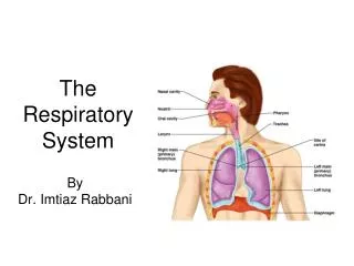
Chapter 22— The Respiratory System
Chapter 22— The Respiratory System. Ch. 22 (Respiratory Sys.) Study Guide. Critically read Chapter 22 pp. 864-886 right before 22.3 “Gas Exchange and Transport” section Comprehend Terminology (those in bold)

Chapter 22— The Respiratory System
E N D
Presentation Transcript
Ch. 22 (Respiratory Sys.) Study Guide Critically read Chapter 22 pp. 864-886 right before 22.3 “Gas Exchange and Transport” section Comprehend Terminology (those in bold) Study-- Figure questions, Think About It questions, and Before You Go On (section-ending) questions Do end-of-the-chapter questions: Testing Your Recall— 1-5, 7, 10, 11-18 True or False– 1, 2, 4-6, 8 Testing Your Comprehension– 1, 4, 5 2
Breathe/Breath (1 or 2) Fear less, hope more; Whine less, breathe more; Talk less, say more; Hate less, love more; And all good things are yours. --Swedish proverb
Breathe/Breath (2 of 2) Every day brings a chance for you to draw in a breath, kick off your shoes, and dance. --Oprah Winfrey
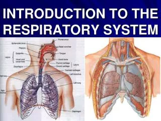
§ I. Anatomical Consideration Self-Check Question: As we breathe in, what respiratory organs, in order, does air pass through? Answer: Nose (mouth) . . . Fig. 22.1
§ Organs of Respiratory System 1 2 3 4 5 6
General Aspects • Airflow in lungs • bronchi bronchioles alveoli • Conducting & Rspiratory (C/R) divisions-- • (C) passages ONLY for airflow, nostrils to bronchioles • (R) distal gas-exchange regions and ________ • Upper/lower (U/L) respiratory tracts • (U) organs in head and neck, nose through larynx • (L) organs of trachea through lungs
§ 1. Nose • Bony and cartilaginous; supported by: • superior half: nasal bones medially and maxillae laterally • inferior half: lateral and alar cartilages • ala nasi: flared portion shaped by alar cartilages and dense CT; forms lateral wall of each nostril • Fig. 22.2 a+b
Conn. tissues shape the nose (Ala nasi)
Nasal Cavity (1) • Extends from nostrils to posterior nares • Vestibule: dilated chamber inside ala nasi (just inside the nostril) • stratified squamous epithelium and vibrissae (guard hairs) • Nasal septum divides cavity into right and left chambers called nasal fossae • Makes up of = Perpendicular plate of ethmoid bone + . . .
Nasal Cavity (2) - Conchae and Meatuses • Superior, middle and inferior nasal conchae • 3 folds of tissue on lateral wall of nasal fossa • mucous membranes lines the cavity • Meatuses: • narrow air passages beneath each conchae • narrowness and turbulence ensures most air contact the mucous membrane. Fig. 22.3
Skull-- Figure 8.4b 1 2 5 6 7 3 4 8 10 9 11
Palatine bones
Functions of the nose • Nose (mouth)—air enters the body through here Functions— • Warm and moisten air • Produce nasal mucus– how much each day? By epi. cells • Cilia– push particles toward the throat
§ 2. Pharynx (throat) Functions— • Common entryway of . . . • Food and air diverge into two separate branches • Air which organ next? • Food which organ next? Which passage way (air or food) is at the anterior? Figure 22.3 b+c 22-17
Lower respiratory tract 22-18
Three Regions of Pharynx Hyoid bone Cricoid cartilage 22-19
§ 2. Pharynx (continued) • Nasopharynx(pseudostratified epithelium) • posterior to choanae, dorsal to soft palate • receives auditory tubes; houses _____ tonsil • 90 downward turn; traps large particles (>10m) • Oropharynx(stratified squamous epithelium) • space between soft palate and root of tongue, inferiorly as far as hyoid bone, contains palatine and lingual tonsils • Laryngopharynx(stratified squamous epi.) • hyoid bone to level of cricoid cartilage
§ 3. Larynx (Voice box) • Anatomy—anterior protrusion called ? • Functions— • Air passageway with cilia • Epiglottis– superior opening of larynx • Voice production by ____________ • Laryngitis—Inflammation of the vocal cords; symptoms? Three major causes?
Larynx • Glottis – vocal cords and opening between them • Epiglottis • flap of tissue that guards glottis, directs food and drink to esophagus • Infant larynx; epiglottis touches soft palate • higher in throat, forms a continuous airway from nasal cavity to the larynx that allows breathing while swallowing • by age 2, more muscular tongue, forces larynx down to lower position
Nine Cartilages of Larynx The superior three (large): • Epiglottic cartilage (1)- most superior • Thyroid cartilage (1)– largest; laryngeal prominence is the Adam’s apple • Cricoid cartilage (1)- connects larynx to trachea • Fig. 22.4
Views of Larynx 1 2 4 5-6 3
Nine Cartilages of Larynx The other 3 small pairs of cartilages: • Arytenoid cartilages (2) - posterior to thyroid cartilage • Corniculatecartilages (2) - attached to arytenoid cartilages like a pair of little horns The above two pairs of cartilages function in speech • Cuneiformcartilages (2) - support soft tissue between arytenoids and epiglottis
Walls of Larynx • Interior wall has 2 muscular folds on each side, from thyroid to arytenoid cartilages • Vestibular folds (superior pair) and vocal cords/folds (inferior) (produce sound) • Intrinsic muscles (deep)- rotate corniculate and arytenoid cartilages (Fig. 22.6) • adducts (tightens: high pitch sound) or abducts (loosens: low pitch sound) vocal cords • Extrinsic muscles (superficial)- connect larynx to hyoid bone, elevate larynx during swallowing
low pitch High pitch
§ 4.Trachea (windpipe) Anatomy/Histology: Beginning of lower respiratory tract (Fig. 22.7 a-c +x) • Rigid tube 5 in. long and 1 in. diameter, anterior/posterior (?) to the esophagus • Supported by 16 to 20 C-shaped rings; openings facing anterior/posterior (?) • The lowermost cartilage called ________ • A smooth m. (trachealis) spans opening in rings, adjusts airflow; facing (ant./post.?) • (Histology) Larynx and trachea lined with ciliated pseudostratified columnar epi. which functions as mucociliary escalator
Larynx See next three slides Trachea Carina
ID structures: Practice at home A B C D E
§ 4. Trachea (continued) Functions: • Air passageway • Warm and moisten air • Remove particles & debris Clinical applications: • Trachea obstruction and Heimlich Maneuver • Tracheostomy (Insight 22.1) when the obstruction is superior to the level of the larynx; pitfall?
§ 5. Bronchi (supported by cartilages) • Primary bronchi (2); with C-shaped rings • from trachea; after 2-3 cm enter hilum of lungs • right bronchus slightly wider and more vertical • Secondary (lobar) bronchi (2 L. lung+ 3 R. lung); one secondary bronchus for each lobe of lung; cartilage plates • Tertiary (segmental) bronchi (8 L. lung + 10 R. lung); cartilage plates • bronchopulmonary segment: portion of lung supplied by each tertiary bronchus Fig. 22.7
§ 6. Bronchioles Bronchioles(lack cartilage; 1 mm or less in diameter; ciliated simple columnar to ciliated simple cuboidal epi.) • Each divides into 50 - 80 terminal bronchioles • Mostly nonciliated simple cuboidal; end of conducting division • Each terminal bronchiole branches into respiratory bronchioles (respiratory div. now); smallest ones are nonciliated epi. • Each divides into 2-10 alveolar ducts (nonciliated simple squamous epi.); end in alveolar sacs • Fig. 22.11
§ Bronchial tree Def. --Highly branched system of air tubes from the primary bronchi to about 65,000 terminal bronchioles Resemble inverted trees Fig. 22.0 + X
CO 22 Right lung; 10 segments Left lung; 8 segments Each broncho-pulmonary segment by a different color of resin
Right lung; 10 broncho-pulmonary segments Left lung; 8 broncho-pulmonary segments
§ 7. Lungs (Fig. 22.9 a + b) • Concave base and blunt apex • Costal surface-- • Concave mediastinal surface— • The hilum (hilus)– slits/depression where bronchi, blood vessels, nerves entering/leaving • The right lung– shorter; the left lung– narrower, with cardiac impression • L– 2 lobes separated by a fissure • R– 3 lobes separated by two fissures
§ 8. Alveoli meaning hollow • Def.– tiny air sacs where . . . • Anatomy/physiology— • Each alveolus– single layer of epithelium surrounded by ____________________ • Numerous alveoli (150 million) in each lung Figure 22.12
§ Alveoli—Pore of Kohn Pore of Kohn • Location? • Function? • Analogy— Fig. x
Smooth muscle 1.Terminal bronchiole A. Branch of pulmonary artery C. Branch of pulmonary vein 2. Respiratory Bronchiole (beginning of respiratory division) B. Pulmonary capillaries 3. Alveolus Alveolar sac Pores of Kohn
§ Alveoli—Three types of cells • Squamous (Type I) alveolar cells— • Location? • Function--Gas exchange through these sites; What type of epi.? • Respiratory membrane– 0.5 micrometer; the barrier between alveolar air and _____ • Great (Type II) alveolar cells— • Location? Embed within alveolar walls • Functions— secretes surfactant & repairs Figure 22.12
Fluid lining With surfactant C. Alveolar macrophage B. Great alveolar cell Alveolus Pulmonary capillary O2 A. Squamous alveolar cell Respiratory mem. RBC
§ Alveoli—Three types of cells • Alveolar macrophages (dust cells)— • Most numerous of all cells in the lung • Large tissue-bound phagocytes • Location– within the alveolar lumen • Function-- Phagocytosis