Urine Analysis
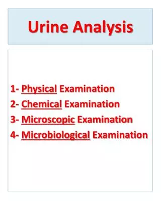
Urine Analysis. 1- Physical Examination 2- Chemical Examination 3- Microscopic Examination 4- Microbiological Examination. Collection of urine. Early morning sample-qualitative Random sample- routine 24hrs sample- quantitative Midstream sample-UTI Post prandial sample-D.M.

Urine Analysis
E N D
Presentation Transcript
Urine Analysis 1- Physical Examination 2- Chemical Examination 3- Microscopic Examination 4- Microbiological Examination
Collection of urine Early morning sample-qualitative Random sample- routine 24hrs sample- quantitative Midstream sample-UTI Post prandial sample-D.M
24 hour urine sample For quantitative estimation of proteins For estimation of vanillyl mandelic acid, 5-hydroxyindole acetic acid, metanephrines For detection of microalbuminuria
Urine Analysis Collection of Urine for Analysis • Urine is collected over a period of 24 hours. • A preservative (as toluene, chloroform, thymol & formalin) is added to prevent contamination of the urine • keeping urine in refrigerator is greatly advisable especially in hot weather.
1 2 3
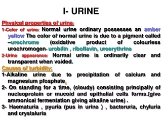
URINE ANALYSISPhysical Examination 1- Volume 2- Specific Gravity 3- Aspect 4- Color 5- Odor 6- Deposit 7- Reaction (pH)
URINE ANALYSISPhysical Examination 1- Volume: Normal urine volume in 24 hours is 600-2000 ml. The night urine is generally < 400 ml. In pregnancy the usual diurnal variation is reversed causing nocturnia & excretion of dilute urine 1-Urine volume increases (Polyuria) in the following conditions: Physiological: • Increased fluid intake • Diuretic Pathological: • Diabetes mellitus (type-1 & type-2) • Diabetes insipidus(due to decrease of ADH) • Chronic renal failure 2- Urine volume decreases (Oliguria or anuria) in the following conditions: • Dehydration • Acute renal failure • Obstruction
URINE ANALYSISPhysical Examination 2- Specific gravity (SG): • Specific gravity measures solute concentration (urea and sodium). • Normally the specific gravity ranges between 1.015-1.025. 1- Increased in • Dehydration (with oliguria) • Diabetes Mellitus (with polyuria) • Acute renal failure (with oliguria) 2- Decreased in • Diabetes insipidus (with polyuria) • Fixed specific gravity (isosthenuria)=1.010 Seen in chronic renal disease when kidney has lost the ability to concentrate or dilute
URINE ANALYSISPhysical Examination 3- Appearance: Normal fresh urine: clear (transparent) Abnormal : Cloudy urine may indicate possible abnormal constituents such as white cells, epithelial cells, crystals and bacteria. N.B. Stored urine with no preservative & no cooling may turn clear urine samples into cloudy. ( d.t. pptn of amorphous crystals, (urates in acidic pH and phosphates in alkaline pH), mucus or growth of bacteria.)
URINE ANALYSISPhysical Examination 4-Color: Normal color: pale yellow (amber yellow) due to the presence of pigments of urobilinor urobilinogen Abnormal colors of urine: • Colorless • Orange • Greenish yellow • Red • Black • Smoky
URINE ANALYSISPhysical Examination Color (cont.) 1- Colorless Urine: • Chronic renal failure • Diabetes insipidus. 2- Orange Urine: • Ingestion of large amount of carotenoids (vitamin A) 3- Yellowish brown urine: due to presence of billirubinin cases of: • Obstructive Jaundice • Hepatic Jaundice
URINE ANALYSISPhysical Examination Color(cont.) 4- Red urine: due to presence of blood,hemoglobin & RBCs. 5- Black urine: • Methemoglobin • Homogentisic acid in alkaptonuria • Malignant malaria (black water fever due to Malaria falciparum). • Melanin(melanoma) 6- Smoky urine: • presence RBCs. in the urine, in cases of acute glomerulonephritis
URINE ANALYSISPhysical Examination 5- Odor: Normal Urineferous odor: The normal odor of fresh voided urine sample Abnormal Odors 1- Fruity odor due to presence of acetone in the urine as in diabetic ketoacidosis 2- Ammonia odor due to release of ammonia as result of: the bacterial action on urea in the contaminated urine or long standing exposed urine samples. 3-Putrid or foul odor : In fresh urine is due to urinary tract infection Other specific abnormal odors are associated with rare disorders of amino acid metabolism 4- Sweet odor : glucose
URINE ANALYSISPhysical Examination 6- Foam • Normal urine will foam slightly when stopped and shaken, and the foam will be white. • The formation of abundant white foam indicates the presence of large concentrations of protein. • A characteristic abundant vivid yellow color foam is shown by urine containing bilirubin
URINE ANALYSISPhysical Examination 7- Deposits: • Normally the urine is devoid of deposits. • The presence of deposits is mainly due to various types of crystals, salts and cells.
URINE ANALYSISPhysical Examination 8- Reaction (pH): Normally: The pH of urine varies from 4.6 - 8.0 1- Acidic urine: • Large intake of meat & certain fruits (cranberries) • Metabolic & respiratory acidosis • Starvation • Severe diarrhea( together with acidosis) 2- Alkaline urine: • Vegetarians • Metabolic & respiratory alkalosis • Urinary tract infection by urea splitting bacteria which spliturea to ammonia (alkaline) • Severe vomiting
Glucose Bilirubin Ketones Specific Gravity Blood pH Protein Urobilinogen Nitrite Leukocyte Esterase Chemical Analysis Urine Dipstick
URINE ANALYSISChemical Examination Normal Constituents of Urine Normal urine contains about 50g of solids dissolved in about 1.5 liters of water per day. Urine contains organic and inorganic solids. A) Chief Inorganic Solids • Sodium, potassium & chlorides • Smaller amounts of calcium, magnesium, sulfate & phosphates • Traces of iron, copper, zinc and iodine. B) Chief Organic Solids: 1- Non-protein nitrogen: • amino acids, ammonia, urea, uric acid , creatine & creatinine 2- Organic acids: • lactic acid, citric acid & oxalic acid • ketone bodies (few amounts) 3- Sugars: • Normally not more than 1g of sugars is excreted in the urine per day. • Sugars cannot be detected by ordinary tests. • Very small amounts of glucose not exceeding 150 mg of glucose are normally excreted per day. • Other sugars present in urine are: pentose and lactose . • Lactosuria occurs in infant and in women during the late months of pregnancy and during lactation
URINE ANALYSISChemical Examination Abnormal Constituents of Urine 1- Proteins (proteinuria) 2- Sugars (glucosuria, fructosuria & galactosuria) 3- ketone Bodies (ketonuria) 4- Billirubin (billirubinuria) & Bile Salts 5- Nitrites
URINE ANALYSISChemical Examination 1- Proteins:(proteinuria) Proteinuria Presence of more than 150 mg proteins in urine in 24 hours detected by ordinary laboratory means heavy proteinuria : >4 gm/24 hours moderate proteinuria: 1 - 4 gm/24 hours minimal proteinuria: < 1.0 gm/24 hours
URINE ANALYSISChemical Examination 1- Proteins: (proteinuria) Proteinuria is divided into prerenal, renal and postrenal proteinuria. 1-Prerenal proteinuria: • Bence-Jones protein: This abnormal gamma globulin (light chains only) is synthesized by malignant plasma cells (multiple myeloma). It precipitates at 60oC, redidssolves at 100oC and reprecipitates on cooling. • Heart failure 2-Renal proteinuria: • Severe muscular exercise • After prolonged standing • Acute glomerulonephritis • Nephrotic syndrome 3-Postrenal proteinuria: • Lower urinary tract inflammation, tumors or stones.
Tests for proteins Test – HEAT & ACETIC ACID TEST Principle-proteins are denatured & coagulated on heating to give white cloud precipitate. Method-take 2/3 of test tube with urine, heat only the upper part keeping lower part as control. Presence of phosphates, carbonates, proteins gives a white cloud formation. Add acetic acid 1-2 drops, if the cloud persists it indicates it protein (acetic acid dissolves the carbonates/phosphates)
URINE ANALYSISChemical Examination Negative Trace (100 mg/dL) + (250 mg/dL) ++ (500 mg/dL) +++ (1000 mg/dL) ++++ (2000+ mg/dL) Glucose Chemical Principle Glucose Oxidase Glucose + 2 H2O+ O2---> Gluconic Acid + 2 H2O2 Horseradish Peroxidase 3 H2O2 + KI ---> KIO3 + 3 H2O Read at 30 seconds
URINE ANALYSISChemical Examination 2- Sugars: (glycosuria) Glucose (Glucosuria); Presence of detectable amount of glucose in urine which occurs in the following conditions: Glycosuria with hyperglycaemia- Uncontrolled Diabetes Mellitus (DM) ,acromegaly, cushing’s disease, hyperthyroidism, drugs like corticosteroids.) - Glycosuria without hyperglycaemia- Renal glucosuria with lowering of renal threshold : e.g. during pregnancy (gestational diabetes). Fructose(Fructosuria): Presence of fructose in urine & may be due to: • - Alimentary causes following the ingestion of large amounts of fructose Fructosemia & herditary fructose intolerance (Metabolic disorders of fructose). Galactose (Galactosuria): Presence of galactose in urine& may be due to: - Alimentary causes following the ingestion of large amount of galactose. - Galactosemia
URINE ANALYSISChemical Examination 3- Ketone Bodies (Ketonuria): Presence of acetone, acetoacetic acid and βhydroxybutyricacid in urine due to: • Diabetic ketoacidosis(uncontrolled DM) • Starvation • Unbalanced diet: high fat & low carbohydrates diet.
URINE ANALYSISChemical Examination 4- Bilirubin (bilirubinuria) Billirubin appears in urine in cases of: • Hepatocellular Jaundice: as in viral hepatitis • Obstructive Jaundice as any cause of obstruction of bile duct
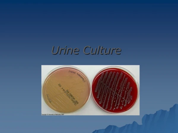
URINE ANALYSISChemical Examination 5-Nitrites • In bacteruria in urine (in cases of Urinary Tract Infection, UTI) • ( nitrate will change to nitrite by some bacteria which are generally gram-negative bacteria)
MICROSCOPIC URINALYSIS a)Crystals :urates andoxalates (acid urine), tripple phosphate (ammonium magnesium phosphate [NH2MgPO4]) (alkaline urine) b)Casts : albuminoid substances released from epithelial c)Parasiticova ; and d)Cells :Pus cells or RBCs.
MICROSCOPIC URINALYSIS • A first morning , midstream, clean catch specimen is generally the specimen of choice. • This specimen is preferred since it is most concentrated and thus small amounts of abnormal constituents are more likely to be detected • Specimen should be centrifuged and examined within 2 hours of collection as RBCs, WBCs and casts disintegrate rapidly especially in dilute ( sp gr.<1010 ) urine of an alkaline pH
Microscopic examination Microscopic urinalysis is done simply pouring the urine sample into a test tube and centrifuging it (spinning it down in a machine) for a few minutes. The top liquid part (the supernatant) is discarded. The solid part left in the bottom of the test tube (the urine sediment) is mixed with the remaining drop of urine in the test tube and one drop is analyzed under a microscope
MICROSCOPIC URINALYSIS 1.Red Blood Cells( N < 3/HPF) Hematuria is the presence of abnormal numbers of red cells in urine due to: a. Glomerular damage b. Tumors c. Urinary tract stones d. Upper and lower urinary tract infections Two Types of Hematuria Gross hematuria means that the blood can be seen by the naked eye. The urine may look pinkish, brownish, or bright red. Microscopic hematuria means that the urine is clear, but blood cells can be seen when urine is looked at under a microscope or tested ina lab. When blood is present in chemical screen , it is important to differentiate between haematuria from hemoglobinuria and myoglobinuria. With haematuria, red blood cells are present /HPF but absent in the others
RBC's may appear normally shaped, • Swollen by dilute urine (in fact, only cell ghosts and free hemoglobin may remain) or crenated by concentrated urinein dilute or hypotonic ( low specific gravity ) • In concentrated urine( high specific gravity) the red cells appear crenated
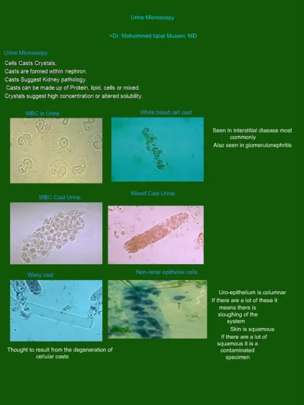
White Blood Cells N < 5/HPF, Pyuriarefers to the presence of abnormal numbers of leukocytes that may appear with infection in either the upper or lower urinary tract or with acute glomerulonephritis. -Repeated sterile cultures in presence of pyuria may indicate: -The patient is on antibiotic therapy -The presence of an organism that does not grow on ordinary media as T.B. -Non- bacterial uretheritis or cystitis as viral infection
Epithelial Cells • Renal tubular epithelial cells, contain a large round or oval nucleus and normally slough into the urine in small numbers. • However, with nephrotic syndrome and in conditions leading to tubular degeneration, the number sloughed is increased
Casts • Urinary casts are formed only in the distal convoluted tubule (DCT) or the collecting duct (distal nephron). • The proximal convoluted tubule (PCT) and loop of Henle are not locations for cast formation. Hyaline casts are composed primarily of a mucoprotein (Tamm-Horsfall protein) secreted by tubule cells. The Tamm-Horsfall protein secretion (green dots) is illustrated in the diagram below, forming a hyaline cast in the collecting duct:
The factors which favor protein cast formation are: *low flow rate, *high salt concentration, *and low pH, all of which favor protein denaturation and precipitation, particularly that of the Tamm-Horsfall protein. Protein casts with long, thin tails formed at the junction of Henle's loop and the distal convoluted tubule are called cylindroids. Hyaline casts can be seen even in healthy patients
Red blood cells may stick together and form red blood cell casts. Such casts are indicative of glomerulonephritis, with leakage of RBC's from glomeruli, or severe tubular damage White blood cell casts are most typical for acute pyelonephritis, but they may also be present with glomerulonephritis. Their presence indicates inflammation of the kidney, because such casts will not form except in the kidney.
Types of casts Acellular casts Hyaline casts Granular casts Waxy casts Fatty casts Pigment casts Crystal casts Cellular casts Red cell casts White cell casts Epithelial cell cast
Hyaline casts The most common type of cast, hyaline casts are solidified Tamm-Horsfall mucoprotein secreted from the tubular epithelial cells Seen in fever, strenuous exercise, damage to the glomerular capillary
Granular casts Granular casts can result either from the breakdown of cellular casts or the inclusion of aggregates of plasma proteins (e.g., albumin) or immunoglobulin light chains indicative of chronic renal disease
Waxy casts waxy casts suggest severe, longstanding kidney disease such as renal failure(end stage renal disease).