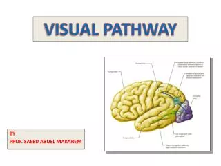
Cranial Nerves: Functions and Components
Explore the intricate system of cranial nerves, their functions, and components. Learn about the unique roles of sensory, motor, and mixed nerves in transmitting signals to and from the brain. Discover how the spinal and cranial nerves differ in their functional components and the specialized functions of special senses. Dive into the organization of motor and sensory nuclei within the brainstem and understand the significance of each in controlling various bodily functions. Unravel the complexities of the cranial nerves and their vital roles in maintaining sensory and motor functions of the body.

Cranial Nerves: Functions and Components
E N D
Presentation Transcript
Cranial Nerves • 12 pairs, (two are attached to the cerebrum and 10 are attached to the brain stem • Nine are attached to the ventral surface of the brain stem, while one is attached to the back of the midbrain (Trochlear). • They leave the cranial cavity by passing through small foramina in the skull bones • Both ‘names’ and ‘numbers’ are used to identify them • Their names indicate either their distribution or their function • Their numbers (ROMAN numerals) indicate the order in which the nerves arise from the brain.
CranialNerves Like all nerves, cranial nerves are made up of bundles of axons Cranial nerves may be sensory, or motor, or mixed, and may contain somatic and/or autonomic fibers. • The olfactory system is closely associated functionally and structurally with the limbic system. • The nuclei of the last 10 cranial nerves are arranged in 7 columns in each side of the middle line in the brain stem. • In the brain stem, in each side of the middle line, there are medial3 column contain motor nucleiand the lateral 4 column contain sensory nuclei.
Ventral surface of brainstem showing attachment of the cranial nerves Trochlear nerve s the only nerve that rise from the dorsal surface of the midbrain
Olfactory Optic Occulomotor Trigeminal Trochlear Abducent Facial & Vestibulo- cochlear Glossopharyngeal Vagus Hypoglossal Accessory
Functional Components of The Spinal Nerves • General Somatic Afferent (GSA): Carries information from skin, muscles, ligament and joints • General Somatic Efferent (GSE): Activate skeletal muscles • General Visceral Afferent (GVA): Carries sensory information from visceral organs • General Visceral Efferent (GVE): Activate visceral (smooth) muscles and glands All the spinal nerves carry all the four components
Spinal Nerves Functional Components • General somatic afferent (GSA) • General somatic efferent (GSE) • General visceral afferent (GVA) • General visceral efferent (GVE) NB. All the spinal nerves carry all the four components
Functional Components of The Cranial Nerves • Four like the spinal nerves (GSA, GSE, GVA, GVE) • Three additional: • 1-Special Somatic Afferents (SSA): • Mediate special sensations of vision and hearing & equilibrium (2&8) • 2-Special Visceral Afferents (SVA): • Mediate visceral sensation of taste & olfaction • 3- Special Visceral Efferent (SVE): • Activate muscles of face, palate, mouth, pharynx and larynx (derivatives of branchial arches) Not all the cranial nerves carry all the components
Cranial Nerves Functional Components • Four like the spinal nerves • General somatic afferent (GSA) • General somatic efferent (GSE) • General visceral afferent (GVA) • General visceral efferent (GVE) • Three additional: (for special senses & pharyngeal arch muscles) • Special somatic afferents (SSA): vision & hearing • Special visceral afferents (SVA): taste & olfaction • Special visceral efferent (SVE): supplying muscles derived from pharyngeal arches NB. Not all the cranial nerves carry all the components
Motor Nuclei column • I-General Somatic Efferent (GSE), column: • Motor nuclei which are in line with the anterior horn cells of the spinal cord. • It give rise to motor fibers which supply striated muscles developed from the myotomes. (3,4,6 &12), (eye & tongue). • These are: • 1- Hypoglossal nucleus in the medulla oblongata. • 2- Abducent nucleus in the caudal Pons. • 3- Oculomotor and Trochlear nuclei in the midbrain.
Motor Nuclei Column • II- General Visceral Efferent (GVE), column: motor nuclei which give parasympatheticfibers to supply smooth muscles of the organs, blood vessels and glands.(3,7,9 &10) • It is homologues to the lateralhorn cells in the spinal cord. • These are: • 1- Dorsal motor nucleus of vagus • 2- Inferior salivatory nucleus. • 3- Superior salivatory nucleus. • 4- Edinger-Westphal nucleus.
Motor Nuclei Column • III- Special Visceral Efferent (SVE) Column: • Motor nuclei which supply muscles developed from the pharyngeal arches, (Branchiomotor cell column). • These are (5,7,9 &10) • 1-Ambiguus nucleus, in medulla (9, 10 & cranial part of accessory nerve) • 2-Motor nucleus of the trigeminal nerve, in the mid Pons • 3-Motor nucleus of the facial, nerve in the caudal Pons.
Sensory Nuclei Column • I-General Visceral Afferent (GVA) column: Sensory nucleus which receive, visceral afferent from pharynx, larynx, esophagus ,thoracic and abdominal viscera: Nucleus solitarius. ----------------------------------------- II- Special Visceral Afferent ( SVA) column: Sensory nucleus which receives taste sensation: Nucleus solitarius.
Sensory Nuclei Column III- General Somatic Afferent (GSA) column: Sensory nuclei which receive: General sensation: (PTPT) pain, temperature, pressure and touch) from the head): Trigeminal sensory nucleus (Spinal and main sensory nuclei of trigeminal nerve. Deep (proprioceptive) sensation from muscles of mastication and tendons & ligaments of TMJ: Mesencephalic nucleus of the trigeminal nerve ----------------------------------------- IV- Special Somatic Afferent (SSA) column: It receive special sensation from the ear, Vestibulocochlear nuclei.
Sensory Pathway Three Neurons: • First order neuron: • From receptors to sensory nucleus. • (The cell bodies lie in the sensory ganglia) • Second order neurons: • From sensory nucleus to the contralateral thalamus • Third order neurons: • From thalamus to cerebral cortex
Motor Pathway Two neurons • Upper motor neuron: • From cerebral cortex to the motor nucleus, mostly bilateral projection except part of 7th and 12th nerves • Lower motor neurons: • From the motor nucleus to the muscles
Olfactory • Type: • Special sensory • Function: • Smell • Lesion : • Leads to loss of sense of smell, called anosmia
Optic • Type: • Special sensory • Function: • Vision • Lesion: • Leads to visual field defects and loss of visual acuity. • A defect of vision is called anopsia
Occulomotor • Type: • Motor & Parasympathetic • 1- OculomotorNucleus • lies close to the apex of the periaqueductal grey mater of the midbrain. • It lies at the level of the superior colliculus. • Its efferent fibers run in the 3rd CN to LPS and all extraocular muscles except LR6 & SO4. • It emerges in the interpedunclar fossa medial to the crus cerebri.
2-Edinger-WestphalNucleus, lies close to the oculomotor nucleus. Gives preganglionic fibers to the ciliary ganglion. Many of the preganglionic fibers traverse the Red Nucleus Postganglionic fibers run in the short ciliary nerve. It supply the constrictor pupillae and ciliary muscles. Function: Elevation of the upper eyelid, Movements of eyeball, Constriction of pupil and Accommodation for near vision
Lesion results in: • Lateral squint • Ptosis • Diplopia • Pupillary dilatation • Loss of accommodation • Impaired downward & outward movement of the eye ball on the damaged side.
TROCHLEAR • Trochlear nucleus • Lies in the periaqueductal grey of the midbrain at the level of the inferior colliculus. • Axons pass dorsally and cross the midline. • It courses around crus cerebri between posterior cerebral and superior cerebellar arteries.
Trochlear • It runs in the lateral wall of the cavernous sinus then to SOF. • It supplies SO4 • It moves the eye downwards and medially. • Lesion: • diplopia, & difficulty in walking downstairs
Trigeminal • Type: • Mixed • Three divisions: • V1:Ophthalmic (sensory) • V2:Maxillary (sensory) • V3: Mandibular (mixed) • Function: • Sensory: • Carries general sensations (pain, touch, temperature, pressure, vibration) from the face & ant. scalp, orbit, nasal and oral cavity, & anterior 2/3 of tongue
TRIGEMINAL NUCLEI • One large sensory ganglion located in the middle cranial fossa, at the apex of petrous temporal bone, its central process go to: • Trigeminal Sensory Nucleus. • It extends all through the brain stem & upper cervical segments • It is formed of 3 subdivisions: • 1-Chief or Main or principal sensory nucleus: • Lies in pontine tegmentum close to the entry of 5th CN. • It receives touch and pressure.
TRIGEMINALNUCLEI • 2-Spinal nucleus & tract of the trigeminal nerve; • Extends caudally in the medulla and upper cervical segments. • It is continuous below the with substantia gelatinosa of Rolando. • It receives pain & temperature sensations. • From the face, scalp, orbit, nasal and oral cavities, and anterior 2/3 of the tongue
TRIGEMINAL NEUCLEI • 3- Mesencephalic Nucleus: extends rostrally into the midbrain. • It carries proprioceptive afferent fibers from the muscles of mastication and from the TMJ. • The cell bodies of all sensations are present in the trigeminal ganglion, Except proprioceptive sensation which lies in the CNS.
Trigeminal lemniscus. • Axons arising from the trigeminal nucleus decussate to form the contralateral trigeminal tract, or lemniscus (2nd order neuron) • This terminates in the contralateral PMVN of thalamus, then to parietal sensory cortex. • The trigeminal nucleus sends fibers to the cerebellum from which, the cerebellum send fibers to the facial nucleus which mediate facial grimacing and eye closure (corneal reflex).
TRIGEMINALNUCLEI • Motor nucleus of trigeminal nerve • It lies in pontine tegmentum medial to the main sensory nucleus. • Fibers runs in the motor root of the trigeminal, then they join the mandibular nerve. • It supply 8 (4+4) muscles which developed from the 1st pharyngeal arch.
Trigeminal • Lesion: • Loss of general sensations in the area of distribution, • Paralysis of the muscles of mastication
Trigeminal Neuralgia • Compression, inflammation or degeneration of the 5th cranial nerve may result in a condition called trigeminal neuralgia or tic douloureux. • This condition is characterized by recurring episodes of intense stabbing , excoriating pain radiating from the angle of the jaw along a branch of trigeminal nerve. • Usually involves maxillary & mandibular nerves, rarely in the ophthalmic division
Abducent • Type: • Motor • Supplies: Lateral rectus. • Function: moves the eye laterally • Lesion: • Medial squint, diplopia, loss of movement of the eye laterally beyond the midpoint.
Abducent Nucleus • Abducent Nucleus • Lies in caudal Pons beneath the floor of the 4th ventricle. • Fibres pass ventrally and emerge from the Ponto-medullary junction between the pyramid and the Pons. • Abducent nerve passes in the lateral wall of the cavernoussinus then through the SOF to supply the lateral rectus
Facial • Type: • Motor,special sensory, parasympathetic • Function: • Motor to muscles of facial expression (2nd pharyngeal arch, lacrimal gland, nasal and oral mucous membrane submandibular & sublingual salivary glands, taste fibers from the anterior 2/3 of the tongue • Lesion: • Bell’s palsy, loss of taste from anterior 2/3 of tongue, loss of Lacrimation and salivation
FACIALNUCLEI • It joins the brain stem in the cerebellopontine angle. • It consists of two roots: • 1- Lateral root (nervous intermedius), contains sensory & parasympathetic fibers. • 2- Medial root contains motor fibers • Sensory fibers, carry taste from the anterior 2/3rd of tongue, floor of mouth and palate, which end solitary nucleus. • It also carry cutaneous sensation from part of external ear, which end in the spinal nucleus of 5th CN
FACIAL NUCLEI • Motor nucleus of facial nerve • It lies in the caudal Pons. • The axons pass dorsally, looping around abducens nucleus beneath the floor of 4th ventricle. • Axons pass in the motor root 7th CN • N.B. Corticobulbar fibers project bilaterally to the upper part of the motor 7th nucleus, and project to the lower part of the nucleus from the opposite side only.
Bell’s Palsy • Damage to facial nerve results in paralysis of facial muscles: Facial (Bell’s palsy);lower motor neuron lesion (whole face affected) • NB. In upper motor neuron lesion (upper face is intact) . • Face is distorted: drooping of lower eyelid, sagging of the angle of the mouth, dribbling of saliva, loss of facial expressions, loss of chewing, blowing, sucking, unable to show teeth or close the eye on affected side
FACIAL NUCLEI • Superior salivatory nucleus. • Lies in the Pons. • Axons run in the nervous intermedius, to the parasympathetic ganglia: • 1- Submandibular ganglion: • Postganglionic fibers pass to submandibular & sublingual salivary gland. • 2- Pterygopalatineganglion: Postganglionic fibers pass to Lacrimal gland, nasal and oral mucous membrane.
Vestibulo-cochlear • Type: • Special sensory • Function: • Vestibular part: conveys impulses associated with balance of body • Cochlear part: conveys impulses associated with hearing • Lesion: loss of hearing, tinnitus, vertigo, dizziness, ataxia and nystagmus
Glossopharyngeal • Type: Motor,Sensory (general & special) parasympathetic • Function: motor to (Stylopharyngeus), & parotid gland, • carries taste fibers from posterior 1/3 of tongue, general sensations from pharynx & palate. • Lesion: • Dysphagia (difficult swallowing), loss of sensation from throat, loss of parotid secretion and loss of taste from posterior 1/3 of the tongue
Vagus • Type: • Motor (+cranial part of accessory nerve), • Sensory (general & special), • Parasympathetic • Function: • supplies visceral muscles, glands of GIT, muscles of the larynx and pharynx, taste buds on the base of tongue, sensations from the viscera Carotid sinus and carotid body
Lesion Leads to: • Loss of gag reflex • Difficulty in swallowing • Loss of sensations from the abdominal viscera • Loss of taste from the base of tongue & epiglottis • Hoarseness or loss of voice • Gastrointestinal dysfunction • Blood pressure anomalies (with CN IX), fatal if both are cut
Accessory • Type: • Motor, • It has two parts: • cranial & spinal • Function: • Cranial part: unites with the vagus and supplies voluntary muscles of larynx, pharynx and esophagus • Spinal part: supplies (sternomastoid & Trapezius) • Lesion: Difficulty in swallowing, and speech. Inability to turn the head, and raise the shoulder.