Anatomy
Anatomy. Organs. Stem. 1-Young dicot stem 2- Monoct stem 3- Old dicot stem. Root. 1- young dicot root 2- monocot root 3- old dicot root. Leaves. Identifying Stems and Roots Diagnostic :. 1- V.B. are of radial type 2- xylem is always exarch ( protoxylem is located

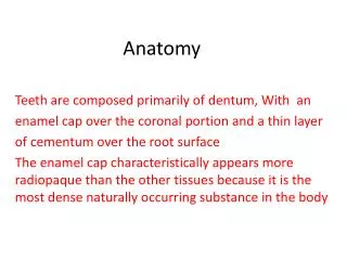
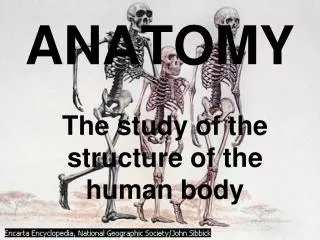
Anatomy
E N D
Presentation Transcript
Organs Stem 1-Young dicot stem 2- Monoct stem 3- Old dicot stem Root 1- young dicot root 2- monocot root 3- old dicot root Leaves
Identifying Stems and Roots Diagnostic: 1- V.B. are of radial type 2- xylem is always exarch ( protoxylem is located near the periphery of the V.B.) 3- Absence of stomata and cuticle Characteristic :
Root anatomy Dermal tissue ( piliferous layer ) One layer , no stomata , no cuticle , the outer wall may extend into root hair Ground tissue Cortex Parenchymatous ( dicot.) , some of them sclerenchyamtous (monocot.) Last layer of cortex termed as Endodermis which characterized by presence of casparian strips { casparian strips : deposits of suberin and lignin on radial wall only Dicot. On radial +inner tangential Monocot .
Endodermis Casparian strips
young dicot root cortex Endodermis phloem Starch Metaxylem protoxylem pith
monocot root epidermis cortex Starch Endodermis phloem Metaxylem protoxylem pith
Old dicot. Root Look at key board
old dicot root Cork (phellem) Cork cambium (phellogen) Phelloderm 2ryphloem Medullary rays Vascular cambium 2ryxylem
Organs Stem 1-Young dicot stem 2- Monoct stem 3- Old dicot stem Root 1- dorsiventral dicot leaves 2- isobilateral dicot leaves 3- monocot leaves 1- young dicot root 2- monocot root 3- old dicot root Leaves
Leaf anatomy Epidermis Upper and lower surface , stomata on both sides Ground tissue ( Mesophyll ) Specialized photosynthetic tissue In Dicot. Divided into palisade Below epidermis and perpendicular to it , contain chloroplasts. spongy tissue Loosely arranged , much less chloroplast than palisade. In Monocot. Mesophyll cells all are alike not diffrantiated into palisade and spongy Vascular tissue Main V.B . In the center ( midrib ) , type of V.B . As the same of stem .
dorsiventral dicot leaves Epidermis Hybodermal layer Palisade Spongy tissue Collenchyma Pericyclic fibers Phloem Metaxylem Protoxylem
Epidermis Palisade Spongy tissue
isobilateral dicot leaves Epidermis Palisade Spongy tissue Collenchyma Pericyclic fibers Phloem xylem
monocot leaves Epidermis Hybodermal layer (sclerenchyma) Ground tissue (parenchyma) Bundle sheath (fibers) Phloem Metaxylem Protoxylem Closed collateral V. B.
Practical work : We will do transverse section in a leaf : tools razors Slides and covers Pith