The Circulatory System
The Circulatory System. Round and round we go!. Your Blood – Fluid Transport. PLASMA!. Plasma is the fluid portion of the blood in which the blood cells move. It is straw-colored and makes up 55% of the total volume of blood!. Red Blood Cells: Oxygen Carriers. Round, disk-shaped cells


The Circulatory System
E N D
Presentation Transcript
The Circulatory System Round and round we go!
PLASMA! • Plasma is the fluid portion of the blood in which the blood cells move. • It is straw-colored and makes up 55% of the total volume of blood!
Red Blood Cells: Oxygen Carriers • Round, disk-shaped cells • Carry oxygen to body cells • Make up 44% of blood • Produced in red bone marrow • Spleen and liver dispose of old red blood cells
How do they carry the oxygen?Hemoglobin to the rescue! • HEMOGLOBIN (iron-containing protein) Oxygen binds to the hemoglobin on red blood cells in the lungs. • Blood passes from the lungs to the body’s cells and where oxygen is needed it is released from the hemoglobin. • Hemoglobin carries some CO2 as well as O2. 70% of this CO2 is made into other substances in the body. 30% travels back to the lungs.
Platelets – The Clotters! • Small cell fragments • Produced from bone marrow • Short life span, living only one week • Help link together a sticky network of protein fibers which forms a scab.
ABO Blood Types • 4 blood types: A, B, AB, O • Differences in blood types are due to presence or absence of antigens • Ex: If you have type A blood, you have the A antigen and the anti-B antibody. If you had a blood transfusion with B blood, your body would attack the new blood cells with the anti-B antibodies! NOT GOOD!
Rh factor in blood • Rhesus factor or Rh factor • Rh is an inherited characteristic • Rh+ if you have Rh antigen; Rh- if not. • Problems occur in pregnant women.
Problems with Rh factor If mother is Rh- and becomes pregnant with Rh+ baby, at birth their blood will mix and the mother will make Rh+ antibodies. If she gets pregnant again with a Rh+ baby, her Rh+ antibodies will destroy the red blood cells in the fetus. Treatment is available to remove the Rh antibodies from her blood so the fetus is not in danger.
Blood Vessels Pathways of Circulation • Arteries – large, thick-walled, muscular, elastic vessels -Carry blood AWAY from heart -Blood is under great pressure
Blood VesselsPathways of Circulation 2. Arterioles – small branches of arteries
Blood VesselsPathways of Circulation • Capillaries – microscopic blood vessels (walls are only 1 cell thick) -Red blood cells move through single file! -Thin capillary walls allow nutrients and gases to diffuse easily between blood cells and surrounding tissues.
Blood VesselsPathways of Circulation • Veins – large blood vessels -Carry blood from tissues TO the heart -Blood is not under great pressure -Blood travels uphill! (legs and arms)
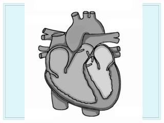
The Heart – The Vital Pump! • The main function of the heart is to keep blood moving constantly throughout the body. • Large organ made of cardiac muscle cells • All mammals have a 4-chambered heart
The Heart – A Vital Pump! • Atria – two upper chambers of heart • Walls are thinner and less muscular
The Heart – A Vital Pump! • Ventricles – two lower chambers of heart • Thicker muscular walls • Performs more work than atria
The Heart – A Vital Pump! • Blood enters the heart through the atria and leaves through the ventricles.
Path of blood through the heart • Both atria fill with blood at the same time. • Right atrium receives oxygen-poor blood from head and body through 2 large veins (vena cava) • Left atrium receives oxygen-rich blood from lungs through 4 pulmonary veins. • Both atria contract pushing blood into the two ventricles.
Path of blood through heart • Both ventricles contract. • When the right ventricle contracts, it pushes oxygen-poor blood out of the heart and toward the lungs through the pulmonary arteries. • When the left ventricle contracts, it pushes oxygen-rich blood our of the heart through the aorta (largest blood vessel in body) to arteries.
Heart Animations Flow of Blood Operation: Heart Transplant High Blood Pressure – Hypertension
Heartbeat – Lub, dub..lub, dub… • Each time the heart beats, a surge of blood flows from the left ventricle into the aorta and into the arteries. This surge is called a pulse! • Heart rate is set by the pacemaker, a bundle of nerves located at the top of the right atrium. The pacemakers send an electrical impulse that tells the atria to contract.
Heartbeat – Lub, dub…lub, dub... • The heart is controlled by the medulla oblongata and the nervous system. • Blood pressure is the force that blood exerts on the blood vessels. • Systolic pressure is when the ventricles contract • Diastolic pressure is when the ventricles relax