The Circulatory System
The Circulatory System. The cardiovascular system is concerned with the transport of blood and lymph through the body Blood circulation may be divided into five major components: the heart, arteries, veins, capillaries and the lymph vascular system


The Circulatory System
E N D
Presentation Transcript
The Circulatory System • The cardiovascular system is concerned with the transport of blood and lymph through the body • Blood circulation may be divided into five major components: the heart, arteries, veins, capillaries and the lymph vascular system • Water and other components of the blood plasma which exude from the blood vessels form the interstitial fluid, which is returned to the circulation by the lymph vascular system • It is customary to divide the circulatory system into the macrovasculature, vessels that are more than 0.1 mm in diameter, and the microvasculature, visible only with a microscope
Tissue Components of the Vascular Wall • The endothelium is a special type of epithelium interposed as a semipermeable barrier between two compartments of the internal medium • Endothelium is highly differentiated to actively mediate and monitor the extensive bidirectional exchange of small molecules and to restrict the transport of some macromolecules • 1. Conversion of angiotensin I to angiotensin II). • 2. Conversion of bradykinin, serotonin, prostaglandins, norepinephrine, thrombin. • 3. Lipolysis of lipoproteins by enzymes located on the surface of endothelial cells, to yield triglycerides and cholesterol • 4. Production of vasoactive factors that affect the vascular tone, such as endothelins, vasoconstrictive agents, and nitric oxide, a relaxing factor.
Vascular Smooth Muscle • Vascular smooth muscle tissue is present in all vessels except capillaries and pericyticvenules. • Smooth muscle cells are frequent and are arranged in helical layers in the tunica media of the blood vessels. • Each muscle cell is enclosed by a basal lamina and by variable amounts of connective tissue both secreted by it. • Vascular smooth muscle cells, mainly of arterioles and small arteries, are frequently connected by communicating (gap) junctions
Vascular Connective Tissue • Components of connective tissue are present in the walls of blood vessels in amounts and proportions that vary based on local functional requirements. • Collagen fibers, a ubiquitous element in the vascular system wall, are found between muscle cells, in adventitia, and in some subendothelial layers. • Collagen types IV, III, and I are present in the basement membranes, tunica media, and adventitia, respectively. • Elastic fibers guarantee the resilient shrinkage of the expanded vascular wall. • Ground substance forms a heterogeneous gel in the extracellular spaces of the vessel wall.
Structural Plan of Blood Vessels • Tunica intima(internal tunic) consisting of : • endothelium (single lining layer of endothelial cells) • sub-endothelial layerinner elastic limiting membrane • Tunica media (middle tunic) consisting of : • circular smooth muscle (or spiral) • concentric elastic lamina (formed by the smooth muscle cells). • Adventitia (outer layer) composed of : • connective tissue surrounding the vessel outer elastic limiting membrane • Vasavasorum:These are small blood vessels supplying oxygen and nutrients to the wall of the artery.
Variations of Vessels • Large Elastic Arteries • Medium (Muscular) Arteries • Arterioles • Metarterioles • Capillaries • PostcapillaryVenules • Small to medium-sized veins • The large veins
Large Elastic Arteries • tunica intima of elastic arteries is thicker than in other arteries. • A layer of loose connective tissue beneath the endothelium (subendothelial connective tissue) allows the tunica intima to move independently from other layers as the elastic arteries distend with the increase in systolic blood pressure. • Distension of the walls is facilitated by concentric fenestrated lamellae of elastic fibres in a thick tunica media. In adult humans, about 40-70elastic lamellae are found in the tunica media of the aorta. • The energy stored in the elastic fibres of the tunica media allows elastic arteries to function as a "pressure reservoir" which forwards blood during ventricular relaxation (diastole). Smooth muscle cells and collagen fibres are present between the layers of elastic fibres. • The external elastic lamina is difficult to discern from other layers of elastic fibres in the tunica media. • The tunica adventitia appears thinner than the tunica media and contains collagen fibres and the cell types typically present in connective tissue
Muscular arteries • The tunica intima is thinner than in elastic arteries. • Subendothelial connective tissue other than the internal elastic lamina is often difficult to discern. • The internal elastic lamina forms a well defined layer. The tunica media is dominated by numerous concentric layers of smooth muscle cells. • the tunica media may contain up to 40 layers of smooth muscle cells • Fine elastic fibres and and a few collagen fibres are also present. The external elastic lamina can be clearly distinguished although it may be incomplete in places. • The thickness and appearance of the tunica adventitia is variable. • The basic structure of the walls of arteries does not change much as we come to the next type of arterial vessels. Size is used to differentiate them from muscular arteries. • Lymphatic capillaries, vasavasorum, and nerves are also found in the adventitia
Arterioles • They are arterial vessels with a diameter below 0.1 - 0.5 mm • Endothelial cells are smaller than in larger arteries, and the nucleus and surrounding cytoplasm may 'bulge' slightly into the lumen of the arteriole. • The endothelium still rests on a internal elastic lamina, which may be incomplete and which is not always well-defined in histological sections. • The tunica media consists of 1-3 concentric layers of smooth muscle cells . It is difficult to identify an external elastic lamina or to distinguish the tunica adventitia from the connective tissue surrounding the vessel • The smooth muscle of arterioles and, to some extent, the smooth muscle of small muscular arteries regulate the blood flow to their target tissues • Arterioles receive both sympathetic and parasympathetic innervation. The final branching of the arterioles finally gives rise to the capillary network
Metarterioles • These are small vessels that are on the border between arterioles and the capillary bed. • They can act as sphincters and cut off the flow of blood into the capillary bed.
Capillaries • The sum of the diameters of all capillaries is significantly larger than that of the aorta, which results in decreases in blood pressure and flow rate. • Their diameter ranges from 4-15 µm. • The wall of a segment of capillary may be formed by a single endothelial cell. This results in a very large surface to volume ratio. The low rate of blood flow and large surface area facilitate the functions of capillaries in: • providing nutrients and oxygen to the surrounding tissue, • and in the excretion of waste products from the body. • Only the tunica intima is present, which typically only consists of the endothelium, its basal lamina and an incomplete layer of cells surrounding the capillary, the pericytes . • Pericytes have contractile properties and can regulate blood flow in capillaries. In the course of vascular remodelling and repair, they can also differentiate into endothelial and smooth muscle cells
There are three different types of capillaries, however the differences are only visible at the ultrastructural level (by light microscopy these differences are not detectable) : • Continuous capillaries • Fenestrated capillaries • Sinusoids • The exchange of materials through capillary walls can be: transcellularvia: • micropinocytotic vesicles in the endothelium (as in continuous capillaries) • fenestrations (as in fenestrated endothelium or sinusoids) • or intercellular via : • gap junctions • spaces between endothelial cells (as in sinusoids of spleen, liver)
Postcapillary Venules • They are larger than capillaries. Small venules are surrounded by pericytes. • A few smooth muscle cells may surround larger venules. • ranging in diameter from 0.1 to 0.5 mm and in length from 0.5 to 70 mm
Small to medium-sized veins • contain bands of smooth muscle in the tunica media. • The tunica adventitia is well developed. In some veins the tunica adventitia contains longitudinally oriented bundles of smooth muscle. • Aside from most veins in the head and neck, small to medium-sized veins are also characterised by the presence of valves. • The valves are formed by loose, pocket-shaped folds of the tunica intima, which extend into the lumen of the vein. The opening of the pocket will point into the direction of blood flow towards the heart. One to three (usually two) pockets form the valve. Blood flowing towards heart will pass the pockets.
The large veins • contain some subendothelial connective tissue in the tunica intima, but both it and the tunica media are still comparatively thin. Collagen and elastic fibres are present in the tunica media. • The tunica adventitia is very wide, and it usually contains bundles of longitudinal smooth muscle. The transition from the tunica adventitia to the surrounding connective tissue is gradual. Valves are absent. • Vasavasorum are more frequent in the walls of large veins than in that of the corresponding arteries - probably because of the lower oxygen tension in the blood contained within them.
Additional Specialisations of Vessels Arteriovenousanastomoses • These represent direct connections between arterioles and venules. • When there is no need for blood flow in the capillary bed these permit direct blood passage (arterial-venous-shunt). Arteriovenousanastomoses are very common in the dermis of the skin. Carotid Bodies • encountered near the bifurcation of the common carotid artery, are chemoreceptors sensitive to carbon dioxide and oxygen concentrations in the blood. • Aortic bodies located on the arch of the aorta are similar in structure to carotid bodies and are believed to have a similar function. Carotid Sinuses • Carotid sinuses are slight dilatations of the internal carotid arteries. • These sinuses contain baroreceptors that detect changes in blood pressure and relay the information to the central nervous system. • The arterial media layer of the sinus is thinner to allow it to respond to changes in blood pressure. The intima and the adventitia are very rich in nerve endings.
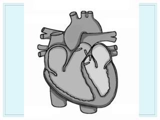
Heart • Its walls consist of three tunics: the internal, or endocardium; the middle, or myocardium; and the external, or pericardium. • The fibrous central region of the heart called the fibrous skeleton, serves as the base of the valves as well as the site of origin and insertion of the cardiac muscle cells. • The endocardium is homologous with the intima of blood vessels. It consists of a single layer of squamous endothelial cells resting on a thin subendothelial layer of loose connective tissue that contains elastic and collagen fibers as well as some smooth muscle cells. • The myocardium is the thickest of the tunics of the heart and consists of cardiac muscle cells • The heart is covered externally by simple squamous epithelium (mesothelium) supported by a thin layer of connective tissue that constitutes the epicardium. • The epicardium corresponds to the visceral layer of the pericardium, the serous membrane in which the heart lies.
The heart has a specialized system to generate a rhythmic stimulus that is spread to the entire myocardium. This system consists of two nodes located in the atriumuam the sinoatrial node and the atrioventricular node, and the atrioventricular bundle. • The atrioventricular bundle is formed by cells similar to those of the atrioventricular node. Distally, however, these cells become larger than ordinary cardiac muscle cells and acquire a distinctive appearance. These so-called Purkinje cells • Both the parasympathetic and sympathetic divisions of the autonomic system contribute to innervation of the heart
LYMPH VASCULAR SYSTEM Functions of the Lymph Vascular System • The lymph vessels return to the blood extracellular fluid from connective tissue spaces. This system ensures the return of water, electrolytes and plasma proteins to the blood. • The lymph vascular system plays a key role in homeostasis of the volume of extracellular fluid. • The lymph vascular system also returns lymphocytes from the lymph nodes to the blood. • The system also transports immunoglobulins (antibodies) from the lymphnodes to the blood
Lymph capillaries • Lymph is a fairly clear, transparent fluid that flows (passively) in small lymph capillaries. These are found in most organs close to blood capillaries (an exception is the CNS). The lymph capillaries begin as small blind-ending tubes. • Typical lymph capillaries have a diameter of 10-50um only. Lymph vessels have very thin walls. The wall of the lymph capillary is composed of a single layer of endothelium (about 0.3um thick). • Lymph capillaries (unlike endothelial cells of blood vessels) lack a basal lamina. • No pericytes or adventitial cells afound. • The lumen is usually free of cells
Lymph ducts • contain one or two layers of smooth muscle cells in their wall (layer the tunica media of lymph vessels). • They also form valves. • Peristaltic contractions of the smooth muscle contribute to the movement of lymph towards the heart in addition to the compression of the ducts by surrounding tissues. • The largest lymph duct of the body, the thoracic duct, drains lymph from the lower half and upper left quadrant of the body and empties the lymph into the circulation by merging with the vascular system close to the junction of the left internal jugular and subclavian veins. • right lymphatic ductempty into the confluence of the right subclavian vein and the right internal jugular vein