Respiratory Function Tests RFTs
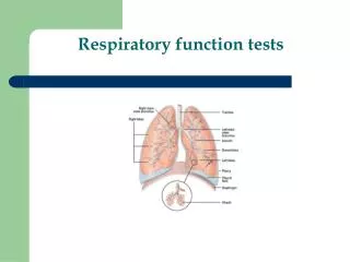
Respiratory Function Tests RFTs. Review Of Anatomy & physiology . Lungs comprised of Airways Alveoli. Airways. Conducting zone : no gas exchange occurs (Anatomic dead space) Transitional zone : alveoli appear, but are not great in number Respiratory zone : contain the alveolar sacs.


Respiratory Function Tests RFTs
E N D
Presentation Transcript
Review Of Anatomy & physiology • Lungs comprised of • Airways • Alveoli
Airways • Conducting zone: no gas exchange occurs (Anatomic dead space) • Transitional zone: alveoli appear, but are not great in number • Respiratory zone: contain the alveolar sacs
The Alveoli • There are approximately 300 million alveoli in each lung. • Their total surface area is 40-80 m2
Mechanics of Breathing • Inspiration • Active process caused mainly by contraction of diaphragm . Accessory muscles may used during exercise and distress • Expiration • Quiet breathing is a passive process but can become active , with forced expiration
Lung Volumes • 4 Volumes • 4 Capacities • Sum of 2 or more lung volumes IRV IC VC TLC TV ERV FRC RV RV
Tidal Volume (TV) • Volume of air inspired or expired during normal quiet breathing • TV = 500 ml IRV IC VC TLC TV ERV FRC RV RV
The extra volume of air that can be inspired over and above the normal tidal volume , when person inspires with full force IRV= 3000 ml The Inspiratory Reserve Volume IRV IRV IC VC TLC TV ERV FRC RV RV
The extra volume of air that can be exhaled over normal tidal volume when person expires forcefully ERV= 1100ml Expiratory Reserve Volume (ERV) IRV IC VC TLC TV ERV FRC RV RV
Volume of air remaining in the lungs at the end of maximum expiration. RV =1200 ml Residual Volume (RV) IRV IC VC TLC TV ERV FRC RV RV
Vital Capacity (VC) • The maximum amount of air a person can expel from the lungs after filling the lungs to their maximum extent and then expires to the maximum extent. Also called Forced vital capacity FVC • VC=4600ml • VC=IRV+TV+ERV IRV IC VC TLC TV ERV FRC RV RV
Inspiratory Capacity (IC) • The amount of air a person can breathe in beginning at the normal expiratory level and distending the lung to the maximum amount. • IC = IRV + TV • IC= 3500ml IRV IC VC TLC TV ERV FRC RV RV
Functional Residual Capacity (FRC) • Volume of air remaining in the lungs at the end of a normal expiration • FRC = ERV + RV • FRC= 2300 ml IRV IC VC TLC TV ERV FRC RV RV
Total Lung Capacity (TLC) • Volume of air in the lungs after a maximum inspiration • TLC = IRV + TV + ERV + RV • =5800ml IRV IC VC TLC TV ERV FRC RV RV
Factors affecting lung volume • Age • Sex • Height • Weight • Race • Disease
CLINICAL SIGNIFICANCE • VC% < 80% is abnormal • RV/TLC% (residual air rate) normal : < 35% emphysema: > 40 % old person can be 50%. • FRC ↑ : emphysema • FRC ↓ : interstitial pulmonary fibrosis
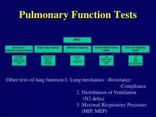
Value of Respiratory function tests • Evaluates 1 or more major aspects of the respiratory system • Lung volumes • Airway function • Gas exchange
Indications • Detect disease • Evaluate extent and monitor course of disease • Evaluate treatment • Measure effects of exposures • Assess risk for surgical procedures
PFTs • Arterial blood gases • Blood PH • Pulse oximeter • Peak flow meter measuring peaked expiratory flow rate. • Spirometry
Peak flow meter measuring peaked expiratory flow rate PEFR • This is extremely simple and cheap test • It describes maximal airflow rate in a given time. • It measures the airflow through the bronchi and thus the degree of obstruction in the airways. • Is best for monitoring the progression of disease
Cont….. • it can detect airway narrowing, commonly used in asthma, Even by the patient himself to know when he need an emergency interference. • the effectiveness of a person's asthma management and treatment plan. • when to stop or add medication, as directed by physician. • what triggers the asthma attack (such as exercise-induced asthma )
To perform this test • Loosen any tight clothing that might restrict your breathing. • Sit up straight or stand while performing the tests • Breathe in as deeply as possible. • Mouthpiece is placed in mouth with lip sealed to prevent escape of air • Blow into the instrument's mouthpiece as hard and fast as possible. • Do this three times, and record the highest flow rate.
Normal values vary based on a person's age, sex, and size • Normal person can empty their chest from full inspiration in 4 sec or less • Prolongation to more than 6 sec indicates airflow obstruction • A fall in peak flow can signal the onset of a lung disease flare, especially when it occurs with symptoms such as: Shortness of breath Increased cough Wheezing
SPIROMETRY • Simple, office-based • Measures flow, volumes • Volume vs. Time • Can determine: - Forced expiratory volume in one second (FEV1) - Forced vital capacity (FVC) - FEV1/FVC
Old version • spirometer bell • kymograph pen • New version • portable
Indications of Spirometry:diagnostic and prognostic • Evaluation of signs and symptoms of pulmonary diseases like asthma and COPD • Screening at-risk populations male smokers >45 years • Monitoring pulmonary drug toxicity • Preoperative assessment • Assess severity of diseases • Follow up response to therapy • Determine further treatment goals • Referral for surgery • Disability
What information does a spirometer yield? • A spirometer can be used to measure the following: • FVC and its derivatives (such as FEV1, FEF 25-75%) • Forced Inspiratory vital capacity (FIVC) • Peak expiratory flow rate • Maximum voluntary ventilation (MVV) • Slow VC • IC, IRV, and ERV • Pre and post bronchodilator studies
Terminology • Forced vital capacity (FVC): • Total volume of air that can be exhaled forcefully from TLC • The majority of FVC can be exhaled in <3 seconds in normal people, but often is much more prolonged in obstructive diseases • Measured in liters (L)
FVC • Interpretation of % predicted: • 80-120% Normal • 70-79% Mild reduction • 50%-69% Moderate reduction • <50% Severe reduction
FEV1 • Forced expiratory volume in 1 second: (FEV1) • Volume of air forcefully expired from full inflation (TLC) in the first second • Measured in liters (L) • Normal people can exhale more than 75-80% of their FVC in the first second; thus the FEV1/FVC can be utilized to characterize lung disease
FEV1 • Interpretation of % predicted: • >75% Normal • 60%-75% Mild obstruction • 50-59% Moderate obstruction • <49% Severe obstruction
Technique • Have patient seated comfortably • Closed-circuit technique • Place nose clip on • Have patient breathe on mouthpiece • Have patient take a deep breath • Blow out the air as fast as possible and as hard and long as possible
Obstructive Disorders Characterized by a limitation of expiratory airflow so that airways cannot empty as rapidly compared to normal (such as through narrowed airways from bronchospasm, inflammation, etc.) Examples: Asthma Emphysema Cystic Fibrosis Restrictive Disorders Characterized by reduced lung volumes/decreased lung compliance Examples: Interstitial Fibrosis Scoliosis Obesity Lung Resection Neuromuscular diseases Cystic Fibrosis ive Vs Restrictive Defect
Obstructive Disorders • Characterized by a limitation of expiratory airflow • Decreased: FEV1, FEV1/FVC ratio (<0.8) • Increased or Normal: TLC
Spirometry in Obstructive Disease • Slow rise in upstroke • May not reach plateau
Restrictive Lung Disease • Characterized by diminished lung volume • Decreased TLC, FVC • Normal FEV1 • Normal or increased: FEV1/FVC ratio
Restrictive Disease • Rapid upstroke as in normal Spirometry • Plateau volume is low
Bronchial Dilation Test • Method: to determine FEV1 and FEV1/FVC% before and after ß2-agonist inhalation • Result: improved rate = after-before ×100% before Positive: >15% • Reversible limitation: asthma