Download
1 / 30
300 likes | 512 Vues
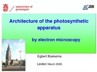
Architecture of the photosynthetic apparatus by electron microscopy. Egbert Boekema Leiden March 2009. Dear keynote speakers in our Solar Biofuels of Microorganisms Workshop

E N D
Architecture of the photosynthetic apparatus by electron microscopy Egbert Boekema Leiden March 2009
Dear keynote speakers in our Solar Biofuels of Microorganisms Workshop <http://www.lorentzcenter.nl/lc/web/2009/333/info.php3?wsid=333>,The workshop is embedded in the Leiden University Honours programme, and there will be 20 of our best bachelor students participating. We havecomfortable slots for the talks and the discussion, and with this email I would like to ask you not to hesitate to include an educational dimension inyour lecture, it will be appreciated, both by our students and by the participant out side your own field in this multidisciplinary workshop.Thanking you for your efforts, and looking forward to seeing you soon in Leiden.Kind regards,-on behalf of the organizers-Corrie Kuster
Unfiltered image of a copper phtalocyanin crystal Electron microscopy is possible at atomic resolution
Photosystem I trimer + 18antenna proteins removal of noise by averaging of many images “single particle averaging” antenna protein = IsiA, the iron stress induced protein A of 37 kDa
main steps in single particle averaging • EM + selection of particle projections • alignment of randomly oriented projections rotational + translational shifts • sorting of projections • statistical analysis + classification • calculation of two-dimensional projection maps • summing of projections into “classes” • calculation of 3D structures symmetrical class tilted class tilted class
Resolution in single particle cryo-3D reconstructions object mass symmetry resolution (Å) number projections 25 17 10 4,000 20,000 75,000 70 s ribosome 3000 kDa none worm hemoglobin 1,000 13 12-fold 4000 kDa Transferrin receptor - transferrin complex 290 kDa 2-fold 36,000 7.5 14 2300 kDa 4-fold 22,000 Ca release channel 823 kDa 14-fold 10,000 GroEL 8 40,000 6 protein 6 rotavirus >5000 kDa >20-fold 8,400 4
First protein at atomic resolution viral protein 6 in rotavirus DLP: 8,400 particles 8,400 x 60 x 13 = 6.6 million copies Zhang et al. PNAS 2008, 105, 1867 Cryo-EM image Electron density map plus amino acid side chain fit (blue wires) lower resolution presentation of virus reconstruction Cryo-EM picture showing Virus particles in a thin layer of ice of a holey carbon film Assignment of amino acid side chains in the 3D map
A test object: worm hemoglobin 12 x 12 proteins 18 linker proteins 18 linker proteins 100 Å
Most complicated step in single particle averaging sorting of projections statistical analysis + classification symmetrical class tilted class tilted class
Classification map after statistical analysis Factor 2 Factor 1 Each dot is a particle in side-view position close in space = high similarity
Classification of aligned side-view projections C D A B G E H I F partition of data set into 9 classes
D I H C G B F E A Position of classes in the classification map
beam Relationship between side-views and top views predominant position 1 “broad type” side view Support film “narrow type” side view predominant position 2 Support film
Sinograms of individual hemoglobin classes to find searchingcommon lines
Worm Hemoglobin 3D Model EM X-ray
sum of 1024 particles 1988 2009 11 Å resolution in negative stain > 2010 Atomic resolution electron counters (800,000 €) 500,000 particles 10000 particles / minute CCD cameras (200,000 €) 50,000 particles 1000 particles / minute Photographic emulsion 5000 particles 1 minute / particle Semi-automation remote-control Handcraft
Seeing is believing The skull from Dali
Complex III (Cytochrome reductase) EM (18 Å resolution) X-ray Seeing is believing The skull from Dali
Example of combining EM and X-ray diffraction Cytochrome reductase – and cytochrome oxidase supercomplex (Heinemeyeret al. 2007 J. Biol. Chem. 282, 12240 maps of the supercomplex and a fragment (left) show enough fine structure to dock the complex III and IV crystal structures accurately into the EM density maps Conclusion: from 15 Å EM data + X-ray structures we get a pseudo-atomic model, which has enough resolution to predict interactionof alpha helicesof different subunits
Scheme of the cyanobacterial membrane Phycobilisome ATPase PSII Cytb6f PSI NDH-1 Cyanobacteria do not have a membrane-bound antenna with LHCH2 Rows of PSII are a scaffold for the phycobilisomes but nobody knows how
Phycobilisome (PBS) PBS components known at high resolution Phycobilisomes are floppy: Structure work on truncated PBSs Need for solving interaction with PSII-PSI, FNR, quenching proteins
Photosystem 1 Complex I ATPase Photosystem 2 Single particle electron microscopy digitonin-solubilized cyanobacterial membranes 50 nm
Selected gallery of projection maps from 15,000 projections Performed on Synechocystis 6803 / Thermocynechococcus elongatus 312 733 351 304 512 512 1230 218 7 50 ~300 291
Phycobilisome fragment Glutamine synthase T-shaped particle ATP synthase GroEl-GroES from the PDB site “molecule of the month displays” Seeing is believing Some ’’’assignments’’’
Small Photosystem II arrays in solubilized membranes from Synechocystis 6803
Analysis of Photosystem II arrays and double dimers 16.7 nm 12.5 nm Phycobilisome model
Analysis of Photosystem II double dimers Double dimer model Is there a specific subunit involved in double dimer formation?