LIVER CIRRHOSIS
680 likes | 1.47k Vues
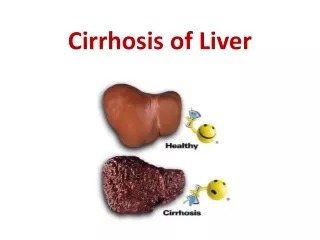
LIVER CIRRHOSIS. THE ANATOMY OF THE PORTAL VENOUS SYSTEM. LIVER CIRRHOSIS. Diffuse disorganization of normal hepatic structure by regenerative nodules that are surrounded by fibrotic tissue. NORMAL. Liver Functions. Metabolism – Carbohydrate, Fat & Protein

LIVER CIRRHOSIS
E N D
Presentation Transcript
LIVER CIRRHOSIS • Diffuse disorganization of normal hepatic structure by regenerative nodules that are surrounded by fibrotic tissue.
NORMAL Liver Functions Metabolism – Carbohydrate, Fat & Protein Secretory – bile, Bile acids, salts & pigments Excretory – Bilirubin, drugs, toxins Synthesis – Albumin, coagulation factors Storage – Vitamins, carbohydrates etc. Detoxification – toxins, ammonia, etc.
LIVER CIRRHOSISCLASSIFICATION (etiologic) • Viral • Alcoholic • Toxic • Autoimmune • Metabolic • Сongestive • Biliary • Cryptogenic
LIVER CIRRHOSISСOMPLICATIONS • HEPATIC ENCEPHALOPATHY • VARICEAL BLEEDING • ASCITES • HEPATORENAL SYNDROME • PORTAL VEIN TROMBOSIS • BACTERIAL PERITONITIS • HEPATOCARCINOMA
LIVER CIRRHOSISCLINICAL SYNDROMES • Astenic syndrome • Pain syndrome • Dyspeptic syndrome • Cholestatic syndrome • Syndrome of jaundice • Portal Hypertension
Spider naevus in liver cirrhosis in the ventral side of theleft shoulder
Xanthelasmas in biliary cirrhosis as a result of primarybiliary cholangitis
Vein dilatation in the abdominal wall of a cirrhotic patient suffering from ascites and jaundice
Clinical Features Hepatocellular failure. Malnutrition, low albumin & clotting factors, bleeding. Hepatic encephalopathy. Portal hypertension. Ascites, Portal systemic shunts, varices, splenomegaly.
Bleeding in Liver disease: vitamin K – in liver gamma-carboxyglutamic acid – for coagulation factors II, VII, IX, and X. Liver disease factor VII is the first to go so the defect will appear initially in the extrinsic pathway, i.e., abnormal PT. When severe it affects both pathways.
Complications: Congestive splenomegaly. Bleeding varices. Hepatocellular failure. Hepatic encephalitis / hepatic coma. Hepatocellular carcinoma.
LIVER CIRRHOSISPLAN of INVESTIGATIONS • Total blood count, Blood group and Rhesus factor • Biochemical analysis (Glucose, Bilirubin, ALT, AST, GGT, Alkaline phosphatase, Albumin, ﻻ-globulins, Cholesterol, Liver tests, Sodium, Potassium, Urea, Creatinine) • Urinanalysis, Diastase of urine • Coagulogram • Markers of Viral Hepatitis(chain polimerase reaction, immunoenzyme analysis) • Immunogram • ECG, Endoscopy, USD, CT, EEG • Diagnostic paracentesis • Needle liver biopsy
Endoscopy reveals large, tortuous esophageal varices thathave a characteristic bluish color
Normal liver with portal vein (VP), inferior vena cava (VC) and right branch of the portal vein (RHA). Subcostal plane: 1 - left branch of VP; 2 - right branch of hepatic vein; 3 - cranial diaphragm
Liver cirrhosis with ascites (longitudinal section): the left lobe of liver is rounded and plump; intrahepatic vessels are reduced. Irregular and inhomogeneous structure. Clear undulatory limitation (arrow) on the underside due to nodular transformation.Wide hypoechoic fringe due to ascites
Post mortem specimen of cirrhotic liver and enlarged spleen Post mortem specimen of banded oesophageal varix
Monitoring of patients with ascites TREATMENT