Cirrhosis liver
250 likes | 1.88k Vues
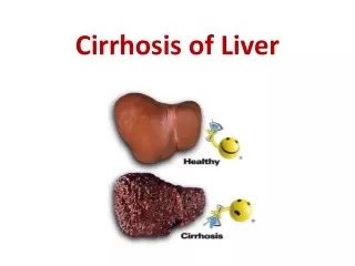
Cirrhosis liver. Dr. Arun R Nair Assistant Professor, dept. of PM. Definition. Cirrhosis, which can be the final stage of any chronic liver disease, is a diffuse process characterized by fibrosis and conversion of normal architecture to structurally abnormal nodules.

Cirrhosis liver
E N D
Presentation Transcript
Cirrhosis liver Dr. Arun R Nair Assistant Professor, dept. of PM.
Definition Cirrhosis, which can be the final stage of any chronic liver disease, is a diffuse process characterized by fibrosis and conversion of normal architecture to structurally abnormal nodules. These “regenerative” nodules lack normal lobular organization and are surrounded by fibrous tissue. The process involves the whole liver and generally is considered irreversible.
CAUSES OF CIRRHOSIS MAIN FACTORS CAUSING CIRRHOSIS Chronic hepatitis C Alcoholic liver disease Nonalcoholic fatty liver disease Chronic hepatitis B Metabolic disorders Hemochromatosis Wilson disease α1-Antitrypsin deficiency Glycogen storage diseases Abetalipoproteinemia Porphyria Hepatic venous outflow obstruction Budd-Chiari syndrome Veno-occlusive disease Right-sided heart failure Drugs and toxins Intestinal bypass Indian childhood cirrhosis OTHER CAUSES OF CIRRHOSIS (<2% OF ALL CASES) Cholestatic and autoimmune liver diseases Primary biliary cirrhosis Primary sclerosing cholangitis Autoimmune hepatitis Intrahepatic or extrahepatic biliary obstruction Mechanical obstruction Biliary atresia Cystic fibrosis
PATHOBIOLOGY & PATHOGENESIS Liver Fibrosis and Cirrhosis
1. The key pathogenic feature underlying liver fibrosis and cirrhosis is activation of hepatic stellate cells. 2. Hepatic stellate cells, which are known as Ito cells or perisinusoidal cells, are located in the space of Disse between hepatocytes and sinusoidal endothelial cells. (vitamin A storage). 3. In response to injury, hepatic stellate cells become activated, as a result of which they lose their vitamin A deposits, proliferate, develop a prominent rough endoplasmic reticulum, and secrete extracellular matrix (collagen types I and III, sulphated proteoglycans, and glycoproteins). 4. They become contractile hepatic myofibroblasts.
5. Liver cells undergo necrosis, the hepatic lobules collapse and this leads to the formation of diffuse fibrous septa. 6. As a compensatory mechanism nodular regeneration of hepatocytes occurs. 7. When the necrosis is associated with collapse of the reticulin framework cirrhosis results. If the reticulin framework is not collapsed but preserved, hepatocytes regrow and reproduce the normal histological pattern. Damage to the reticulin framework results in the formation of abnormal nodules which derive nourishment from the hepatic artery, but without portal and biliary connections. The nodules vary in size from a few millimeters to several centimeters.
10. The liver surface becomes nodular. 11. Hepatic vascular bed is distorted, truncated and obstructed, the obstruction being maximal at the level of sinusoids. Causes several vascular abnormalities ,they are; a. Generalized arterialisation of the liver. b. Formation of shunts between the branches of the hepatic artery, portal vein and hepatic vein. c. Formation of arteriovenous shunts also in the pulmonary circulation. d. Development of a hyperdynamic circulatory state with increased cardiac output and reduced peripheral vascular resistance.
12. Obstruction to portal venous flow results in the development of portal hypertension. 13. As the nodules grow, their centers are rendered ischemic. Once the disease process is initiated, other factors such as autoimmunity, continuing necrosis, and chronic effect of toxins lead to the progression of the pathological lesions. 14. Basement membrane forms in the Disse’s space and this interferes with the metabolic functions of the liver. 15. The necrotic foci stimulate the proliferation of fibroblasts and collagen and fibrous septae develop in the portal zones and hepatic lobules.
Hepatic sinusoid Spleen Porto-systemic collaterals Portal vein Liver Hepatic sinusoid Spleen Portal vein Liver
Morphological Classification • The morphological types are: • a. micronodular, • b. macronodular, and • c. mixed.
Compensated Cirrhosis In this stage, cirrhosis is mostly asymptomatic and is diagnosed either during the evaluation of chronic liver disease or fortuitously during routine physical examination, biochemical testing, imaging for other reasons, endoscopy showing gastroesophageal varices, or abdominal surgery in which a nodular liver is detected. Nonspecific fatigue, decreased libido, or sleep disturbances may be the only complaints.
Decompensated Cirrhosis At this stage, there are signs of decompensation: ascites, variceal hemorrhage, jaundice, hepatic encephalopathy, or any combination of these findings. Ascites, which is the most frequent sign of decompensation, is present in 80% of patients with decompensated cirrhosis
H.C.C C/C Liver Cirrhosis Compensated Cirrhosis Decompensated Cirrhosis Death Complications develops
CLINICAL FEATURES Early stages of the disease are asymptomatic. Vague illhealth, anorexia, loss of weight, loss of libido, impotence, abdominal distention, dependent edema Night blindness (impairment of metabolism of vitamin A). Ascites
In the compensated phase, liver synthetic function is mostly normal, and portal pressure, although increased, is below the threshold level required for the development of varices or ascites. As the disease progresses, portal pressure increases and liver function worsens, thereby resulting in the development of ascites, portal hypertensive gastrointestinal bleeding, encephalopathy, and jaundice. The development of any of these clinically detectable complications marks the transition from a compensated to a decompensated phase. Progression to death may be accelerated by the development of other complications, such as recurrent gastrointestinal bleeding, renal impairment (refractory ascites, hepatorenalsyndrome), hepatopulmonary syndrome, and sepsis (spontaneous bacterial peritonitis). The development of hepatocellular carcinoma may accelerate the course of the disease at any stage
Complications Variceal Hemorrhage Ascites and Hyponatremia Spontaneous Bacterial Peritonitis Hepatic Encephalopathy Pulmonary Complications
DIAGNOSIS • The diagnosis may often require histologic confirmation by liver biopsy, which is the “gold standard” for the diagnosis of cirrhosis. • Physical Examination. • Laboratory Tests- The most sensitive and specific laboratory finding suggestive of cirrhosis in the setting of chronic liver disease is a low platelet count (<150,000/μL), which occurs as a result of portal hypertension and hypersplenism • Imaging Studies Dr. Arun R Nair. Assistant Professor Dept. of PM