Download
1 / 26
310 likes | 1.09k Vues
Cirrhosis of the liver. January 8 th 2013. Cirrhosis of the liver: OUTLINE. The Case Histology Etiology of Cirrhosis Ecology of Cirrhosis The Care of the Cirrhotic patient Back to the Case. THE CASE. 33 YO Female with right upper quadrant abdominal pain. The case.

E N D
Cirrhosis of the liver January 8th 2013
Cirrhosis of the liver: OUTLINE • The Case • Histology • Etiology of Cirrhosis • Ecology of Cirrhosis • The Care of the Cirrhotic patient • Back to the Case
THE CASE • 33 YO Female with right upper quadrant abdominal pain
The case • Liver biopsy revealed: • GRANULOMATOUS HEPATITIS CONSISTENT WITH SARCOIDOSIS • Treatment: • Prednisone for one month • Outcome: • She felt well and returned to work as a professional dancer
What causes cirrhosis? • Repeated insults oxidative damage Kupffer and stellate cell activation production of excess collagen and extracellular matrix • Alcoholism • Chronic viral hepatitis • Autoimmune hepatitis • NASH • Biliary cirrhosis (PBC, PSC, autoimmune cholangiopathy) • Cardiac cirrhosis • Inherited metabolic liver disease (hemochromatosis, Wilson’s disease, AAT deficiency, CF) • Cryptogenic cirrhosis
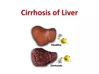
Cirrhotic Liver: Histology • INJURY • DEGENERATION • FIBROSIS • FORMATION OF FIBRO-VASCULAR MEMBRANES • PARENCHYMAL DISSECTION INTO NODULES • REARRANGEMENT OF CIRCULATION • CIRRHOSIS
Effects of cirrhosis: PHYSICAL EXAM • JAUNDICE AND SCLERAL ICTERUS • ASCITES • EDEMA • HEMORRHOIDS • SPLENOMEGALY • FIRM, NODULAR LIVER EDGE • PALMAR ERYTHEMA • SPIDER ANGIOMAS • CAPUT MEDUSAE • PAROTID GLAND ENLARGEMENT • DIGITAL CLUBBING • MUSCLE WASTING • MEN: DECREASED BODY HAIR, GYNECOMASTIA, TESTICULAR ATROPHY • WOMEN: METRORRHAGIA OR AMENORRHEA
EFFECTS OF CIRRHOSIS: LABS • CAN BE COMPLETELY NORMAL IN EARLY COMPENSATED CIRRHOSIS • IN ADVANCED LIVER DISEASE… • ANEMIA: CHRONIC GIB, POOR NUTRITION, HYPERSPLENISM, BONE MARROW SUPPRESION, ZIEVE’S SYNDROME (HEMOLYTIC ANEMIA WITH SPUR CELLS AND ACANTHOCYTES) • THROMBOCYTOPENIA • NORMAL OR ELEVATED TOTAL BILIRUBIN AND ELEVATED DIRECT BILIRUBIN • PROLONGED PT • HYPONATREMIA WITH ASCITES • TRANSAMINITIS
CARE OF THE CIRRHOTIC PATIENT • FACTORS THAT PREDISPOSE CIRRHOTIC PATIENTS TO DECOMPENSATE: • BLEEDING • INFECTION • ALCOHOL INTAKE • MEDICATIONS • DEHYDRATION • CONSTIPATION • OBESITY
Care of the cirrhotic patient: ascites • Ascites is the most common complication of cirrhosis • Caused by Portal Hypertension (hepatic venous pressure gradient >5mmHg) which is caused by.. • Increased intrahepatic resistance • Increased splanchnic blood flow due increased splanchnic lymph • Ascites accumulates when… • Hypoalbuminemia decreased oncotic pressure • Sodium retention perpetuates third spacing • Labs to obtain when performing paracentesis: albumin, protein, cell count and diff, culture, gram stain, AFB, fungal culture, cytology, amylase, lipase, TGs
Care of the cirrhotic patient: ascites • What is SAAG? • Ascites can be managed initially with sodium restriction <2g/d • If moderate ascites, use spironolactone 100-200mg/d and can add furosemide 40-80mg/d • For refractory ascites, consider repeat LVP or TIPS procedure • For repeat LVP, always replace albumin if drain > 5L
Care of the cirrhotic patient: SBP • Spontaneous Bacterial Peritonitis • 25% in-hospital mortality rate • ‘Spontaneous’ due to bacterial translocation • Most common organism: Escherichia coli, and gram positives sometimes found such as Strep viridans, Staph and Enterococcus • If >2 organisms, consider perforated viscus • How do we diagnose SBP? • History, PEX, labs • How do we treat SBP? • 2nd generation cephalosporin • Who should use SBP prophylaxis? • Upper GI Bleed patients • Previous SBP • Ascitic fluid protein < 2.0
Care of the cirrhotic patient: GI bleed • Upper GI bleed secondary to esophageal or gastric varices • PRIMARY PROPHYLAXIS • All patients diagnosed with cirrhosis should have EGD • Increased risk: Red wale sign, hmetocystic spots, cherry-red or white-nipple spots, blue or erythematous • IR can measure gradient between wedge and free hepatic vein; if >12 mmHg at risk for variceal hemorrhage • Nadolol or propanolol • EVL • SECONDARY PROPHYLAXIS: repeat EVL and beta-blockers • ACUTE VARICEAL BLEED TREATMENT • IVF, blood products • Somatostatin • Balloon tamponade • Sclerotherapy • EVL • GAVE: EVL wont work, perform TIPS
Care of the cirrhotic patient: HRS • Hepatorenal Syndrome • Caused by renal vasoconstriction • Low UOP, low urine sodium • 10% patients with advanced cirrhosis, usually those with large ascites • Type 1 versus Type 2 • Treated with midodrine, octreotide, IV albumin • Prognosis for Type 1 is poor unless transplanted
Care of the cirrhotic patient: Encephalopathy • HEPATIC ENCEPHALOPATHY • Caused by gut derived neurotoxins normally removed by liver • Disturbance in diurnal sleep patterns is an early sign • Brain edema can cause herniation • Asterixis, hyperreflexia • Precipitants of HE: hypokalemia, infection, increased protein, GIB, dehydration • TREATMENT: Lactulose, rifaximin, zinc
Care of the cirrhotic patient: Pulmonary complications • HEPATOPULMONARY SYNDROME • Platypnea and orthodeoxia • Triad: liver disease, increased A-a gradient, Intrapulmonary vascular abnormalities • PORTOPULMONARY HYPERTENSION • Pulmonary hypertension in patients with portal hypertension • 2% of patients with cirrhosis • Fatigue, dyspnea, peripheral edema, CP, syncope HEPATIC HYDROTHORAX • PLEURAL FLUID IN A PATIENT WITH ASCITES AND NO CARDIOPULMONARY DISEASE • THROUGH DEFECTS IN DIAPHRAGM • USUALLY R SIDED
CARE OF THE CIRRHOTIC PATIENT: CARDIOMYOPATHY • CIRRHOTIC CARDIOMYOPATHY • Up to 50% of patients with advanced cirrhosis • Normal to increased CO and contractility at rest but blunted response to stress • Diastolic dysfunction • QRS widening • Caused by abnormalities in the β-adrenergic signaling pathway, altered cardiomyocyte membrane fluidity, increased myocardial fibrosis, cardiomyocyte hypertrophy, and ion channel defects • Acute volume overload (TIPS, transplant) or increased demand for CO (infection) can lead to heart failure • Treatment: Beta blockade?
Care of the cirrhotic patient: coagulopathy • Coagulopathy is almost universal in cirrhotic patients • Decreased synthesis of clotting factors and impaired clearance of anticoagulants • Thrombocytopenia • Vitamin K requires biliary excretion for subsequent absorption so this process is diminished • Decreased hepatic mass means decreased synthesis of • Which factors are affected? TREATMENT: IV or IM vitamin K, FFP, platelets
Care of the cirrhotic patient: HCC • Chronic liver disease is the major risk factor for developing HCC. • Most patients with HCC have underlying cirrhosis • Environmental Factors: Food contaminated with aflatoxin and smoking increase risk • Corns, soybeans, peanuts • Diabetes is associated with HCC, and treating with metformin decreases risk • Co-infection with HIV increases risk • SURVEILLANCE: HBV carriers and all patients with cirrhosis • Liver US every 6 mos (+/- AFP level) • If liver nodule found, <1cm get repeat in 3 mos; if >1cm further imaging • TREATMENT: • OLT for patients who meet Milan criteria (single tumor <5cm or <3 tumors each <3cm, no macrovascular invasion • Resection • TACE • Ablation • XRT • Chemotherapy
BACK TO THE CASE • 33 YO F with infiltrative liver disease secondary to sarcoidosis • Initially treated with course of prednisone for one month, did well • 5 mos later developed dyspnea after one flight of stairs stopped smoking • 3 mos after that developed melena and syncope • Finally presented to MGH where she had following workup: • EGD showed varices, gastritis and CT showed hepatosplenomegaly • TTE showed dilated RA + RV and TR • R Heart Cath showed elevated PA pressures not improved with NO • On 6th hospital day after pt developed agitation, AMS, asterixis, worsening SOB she had cardiac arrest and died FINAL DIAGNOSIS: HEPATIC CIRRHOSIS DUE TO END STAGE PRIMARY BILIARY CIRRHOSIS AND PLEXOGENIC PULMONARY HYPERTENSION DUE TO CIRRHOSIS, WHICH CAUSED COR PULMONALE
Take care of me and I’ll take care of you! That’s all folks, Thanks! THE END