Liver Cirrhosis
910 likes | 1.44k Vues
Liver Cirrhosis. Lamya Alnaim, PharmD. Background. Cirrhosis → the end stage of any chronic liver disease. Hepatitis C and alcohol are the main causes Two major syndromes result Portal hypertension Hepatic insufficiency.

Liver Cirrhosis
E N D
Presentation Transcript
Liver Cirrhosis Lamya Alnaim, PharmD
Background • Cirrhosis →the end stage of any chronic liver disease. • Hepatitis C and alcohol are the main causes • Two major syndromes result • Portal hypertension • Hepatic insufficiency. • peripheral and splanchnic vasodilatation with the resulting hyperdynamic circulatory state
Background • Cirrhosis can remain compensated for many years before the development of a decompensating event. • Decompensated cirrhosis is marked by the development of any of the following complications: • Jaundice, • Hemorrhage • Ascites • Encephalopathy. • Other than liver transplantation, there is no specific therapy for this complication.
Background • Other complications occur as a consequence of PHTN and the hyperdynamic circulation. • Gastroesophageal varices result from PHTN, although hyperdynamic circulation contributes • Ascites results from sinusoidal HTN and sodium retention, which is 2ndry to vasodilatation and activation of neurohumoral systems.
Background • The hepatorenal syndrome results from severe peripheral vasodilatation that leads to renal vasoconstriction. • Hepatic encephalopathy is a consequence of shunting of blood through portosystemic collaterals (due to PHTN), brain edema (cerebral vasodilatation), and hepatic insufficiency.
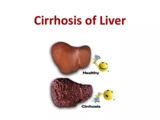
Definition • A chronic disease of the liver with wide spread hepatic parenchymal injury and hepatocyte destruction. • It may lead to anatomic and functional abnormalities of blood vessels and bile ducts
Causes • Alcohol • Viral illness • Biliary dysfunction • Metabolic disorders • Inherited disorders • Drugs • The most common causes are alcoholism and viral hepatitis
Clinical Features • Insidious development • Often produces no clinical manifestations • Common symptoms • Anorexia, nausea, abdominal discomfort, weakness, weight loss, and malaise
Clinical Features • Physical examination: • Enlargement of the liver and spleen due to PHTN • ascities, • peripheral edema, • Jaundice • Spider angiomas • GI bleeding • Palmer erythema • Right upper quadrant pain
Portal Hypertension • Portal vein collects blood from GI tract, pancreas and spleen to the liver • Contains oxygen, nutrients and bacterial waste • A pathway for detoxification and metabolism of absorbed substance. • Fibrosis and nodular regeneration of liver with distortion of hepatic veins is the main cause of ↑ intrahepatic resistance
Portal Hypertension Persistent PHTN lead to • Changes in blood and lymphatic flow → hyperfiltration and ascites • ↑ collateral circulation ↑ the risk for esophageal and gastric varices • Hepatic encephalopathy and hepatorenal syndrome
Lab Findings • Bilirubin> 2mg/dl to 40 mg/dl • AST, ALT, alkaline phosphatase • Aid in early diagnosis, prognosis, and response to treatment • ↑ ALkPo > 3 times normal indicate billiray disease
Lab Findings Albumin • (non-specific protein) & Factor V and VII (specific proteins) can provide information on the functional capacity of the liver • Low albumin < 3 that does not respond to therapy is bad prognosis
Lab Findings • PT • Prolongation due to impaired synthesis of vitamin K dependant clotting factors • No response to VIT K is poor prognosis • BUN • < 5 due to inadequate protein intake and depressed hepatic capacity for urea synthesis • Biopsy • Confirm the presence of cirrhosis
General Management • Largely symptomatic • Maintain fluid and electrolyte balance
General Management • Analgesics: • NSAIDs may worsen gastritis and GI bleeding • Acetaminophen may lead to hepatoxicity • Narcotics may lead to CNS and respiratory depression • Sedatives and hypnotics should be avoided if the patient is in danger of hepatic coma
General Management • Diet • 2000-3000 calorie diet with 1g protein/kg • In encepalopathy, dietary supplentation of BCAAs • Thiamine replacement 50-100mg/da • Iron and folate if patient is anemic • Vitamin K 10 mg sc if PT is elevated, if PT is not improved in 3-5 days D/C
I-Ascities Definition: • Accumulation of protein rich fluid in the peritoneal cavity. • The most common clinical feature Clinical features: • Inability to fit into one's clothes • Abdominal and back pain • Gateroesophageal reflux • SOB secondary to impaired diaphragm movement Or pleural effusions
Ascities Pathophysiology:Underfill theory 1-↑hydrostatic pressure in portal vein and ↓oncotic pressure. • Exudation of fluid from the splanic capillary bed and liver surface when drainage capacity of the lymphatic system is exceeded.
Ascities Pathophysiology:Underfill theory 2-Portal hypertension & ↓ oncotic pressure →↓ arterial blood flow to vital organs → vasoconstriction. • Reduced circulation to kidney activate rennin angiotension sys →↑ aldosterone & Na/ water retention. • Renal K excretion > Na excretion & urinary Na: K ratio abnormal.
Ascities Goals of therapy • Mobilize fluid • Diminish abdominal discomfort, back pain, and difficulty in ambulation • Prevent complications such as bacterial peritonitis, hernias, pleural effusion, hepatorenal syndrome, respiratory distress. • Prevent complications of treatment such as acid-base imbalance, hypokalemia, and volume depletion
Ascities -Treatment A- Sodium Restriction: (500mg-2g/day) • 10-20 mEq/day plus bed rest (to ↓rennin) • Degree of success depends on: • Duration of restriction • Extent of hepatic injury • Patient with urine Na >10 mEq/l likely respond
Ascities -Treatment B-Water Restriction • Effective in dilutional hyponatremia (Na<130) • Patients with low urine sodium <10 mEq/l • Normal renal function • Not effective in • reduced 24-hour Na urinary excretion • Reduced GFR & free water clearance • May lead ↓ renal blood flow and azotemia
Ascities -Treatment C. Diuresis • The cornerstone of treatment • Must be slow • If urinary losses > reabsorption from ascites→ volume depletion , hypotension and renal insufficiency • Should be limited to 0.2-0.3 kg /day in patients without edema • 0.5-1kg /day for patients with edema
Ascities -Treatment I- Spironolactone • An aldosterone-inhibiting agent • Patients have high levels of aldosterone • Increased production • Portal hypertension, ascities, ↓intravascular volume, ↓ renal blood flow activate rennin-ang system • Decreased excretion • Hepatic impairment prolongs half-life due to ↓ metabolism • ↓ albumin↑ unbound hormone in the blood • Dose: 100-200 mg/day, may be ↑ slowly every 2-4 days
Ascities -Treatment I- Spironolactone Monitoring Parameters • weight • urine output • changes in abdominal girth • BUN • Increase in K/Na ratio from pretreatment baseline • Table
Complication of Spironolactone 1-Hypokalemic hyperchloremic metabolic alkalosis and hyponatermia • May occur in untreated cirrhosis • Initial deficiency of K due to diarrhea, vomiting, hyperaldosterone • May be corrected with KCl supplement • Hyponatermia corrected by temporary withdrawal of diuretic and free water restriction 2- Prerenal azotemia • ARF due to overdiuresis with compromise in intravascular volume and decreased renal perfusion • gradual rise in Scr and Bun • If large fluid volume must be removed quickly, paracentesis should be preformed 3- Gynecomastia: • can be related to cirrhosis independent of drug use
Ascities -Treatment II-Other Diuretics • If spironolactone fails to produce diuresis or hyperkalmeia occurs additional diuretic are needed • The dose should be started low 50 mg/day HCTZ or 20-40 mg furosemide and gradually increased • Loop and thiazide diuretics may affect the value of monitoring urinary electrolyte • The may cause excessive sodium loss in the presence of continued hyperaldosteronism
Ascities -TreatmentD- Paracentesis Removal of large amount of ascetic fluid with a needle or catheter Uses • Ascites unresponsive to diuretic therapy • If respiratory and cardiac functions are compromised • Not definitive because fluid quickly reaccumliated due to transudation of fluid from the interstitial and plasma
D- Paracentesis Major complications • 15-100% of the fluid reaccumlates with 24-48 hrs → transient hypovolemia and possibility of shock, encaphalopathy and ARF • Hypotension • Hemconcentration
D- Paracentesis Major complications • Shock • Oliguria • Hepatorenal syndrome • Hemorrhage • Perforation of abdominal vicra • Infection, bacterial peritonitis • Protein depletion
E- Albumin • Combined with paracentesis • Effective as the initial management in tense ascites Typical regimen: Removal of 4-6 l/day with replacement of 40-50g of albumin
E- Albumin Benefits of combination: • More ascetic fluid can be removed • Shorter hospital stay • Superior to diuretic therapy • No worsening of hepatic, renal or CV function • Albumin alone can promote diuresis in ascites & edema by increasing intravascualr volume • The effects are not long lasting • Variceal hemorrhage may be precipitated
F- Dextran 70 • Can be combined with paracentesis • Equally effective to albumin in mobilizing ascities • More cost-effective • Does not correct the underlying hemodynamic abnormalities, so albumin is preferred
G-Surgical therapy 1- Peritoneovenous shunt • An implanted valve in the abdominal wall, with cannula that empties into the vena cava • Urine output as high as 15 L in 24 hrs • Supplemental fruosamide may be needed to prevent vascular overload
G-Surgical therapy 1- Peritoneovenous shunt Contraindications: • Peritonitis, • Recurrent coma • Sever coagulopathy • Significant cardiac failure • Acute alcoholic hepatitis
G-Surgical therapy 1- Peritoneovenous shunt Complication • Pulmonary edema • Coagualopahty • Fever, Wound infection, Septicemia, GI bleeding • Reserved for patients with good renal and hepatic function who fail standard therapies
G-Surgical therapy 2-transjugular intrahepatic portosystemic shunt (TIPS) • a radiologic procedure in which a stent is placed in the middle of the liver to reroute the blood flow. • it makes a tunnel through the liver connecting the portal vein to one of the hepatic veins. • A metal stent is placed in this tunnel to keep the track open.
SBP • SBP is an infection of ascites that occurs in the absence of a contiguous source of infection. • SBP occurs in 10 to 20 % of hospitalized cirrhotic patients. • Early diagnosis is a key issue in the management of SBP. • SBP pathogenesis in patients with cirrhosis is considered • to be the main consequence of bacterial translocation.
SBP-Predisposing factors • Severity of liver disease • Total ascites protein <1 g/dL • GI bleeding • Bacteriuria • Previous SBPRecurrence rates: 43% by 6 mts, 69% by 1 yr and 74% by 2 yrs
SBP-Clinical features • signs may be absent in up to 1/3 • fever/hypothermia • abdominal pain and tenderness • hepatic encephalopathy • diarrhoea • ileus • shock • Unexplained deterioration in a patient with cirrhosis and ascites should lead to diagnostic paracentesis
SBP-diagnosis A diagnostic paracentesis should be performed in • Any patient admitted to the hospital with cirrhosis and ascites, • Any cirrhotic patient who develops compatible symptoms or signs • Any cirrhotic patient with worsening renal or liver function. • Diagnosis is established with an ascites PMN of > 250/mm3
SBP- Treatment • Once an ascites PMN count of >250/mm3 is detected, and before obtaining the results of ascites or blood cultures, antibiotic therapy needs to be started. • The antibiotic that has been most widely used in the treatment of SBP is IV cefotaxime (2g 8 hrly) with which SBP resolves in around 90 90% of treated patients
SBP- Treatment • The combination of amoxicillin and clavulanic acid was shown to be as effective and safe as cefotaxime • Antibiotic treatment can be safely discontinued after the ascites PMN count decreases to below 250/mm3 • duration of antibiotic therapy should be for a minimum of 8 days
Prevention of recurrent SBP • In patients who survive an episode of SBP, the 1-year cumulative recurrence rate is high, at about 70 %. • It is essential that patients be started on antibiotic prophylaxis to prevent recurrence before they are discharged from the hospital.
Prevention of recurrent SBP • Long-term prophylaxis with oral norfloxacin at a dose of 400 mg QD • treatment should be initiated as soon as the course of antibiotics for the acute event is completed. • oral ciprofloxacin at a dose of 250 mg QD could be used, although levofloxacin may be a better alternative given its added gram-positive coverage.
Prevention of recurrent SBP • Weekly administration of quinolones is not recommended given a lower efficacy and an increase in the development of fecal quinolone-resistant organisms. • Prophylaxis should be continuous until disappearance of ascites (i.e., patients with alcoholic hepatitis who stop drinking) or transplant