Download
1 / 103
1.57k likes | 2.43k Vues
Cirrhosis of Liver. Introduction. The term cirrhosis was first used by Rene Laennec (1781-1826) to describe the abnormal liver color of individuals with alcohol induced liver disease. Derived from Greek word Kirrhos means Yellowish brown color. Definition:

E N D
Introduction • The term cirrhosis was first used by Rene Laennec (1781-1826) to describe the abnormal liver color of individuals with alcohol induced liver disease. • Derived from Greek word Kirrhos means Yellowish brown color.
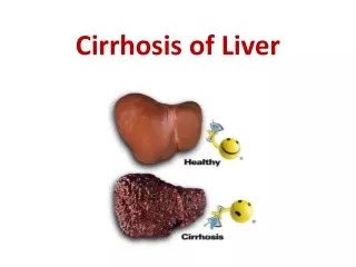
Definition: • Cirrhosis is a chronic progressive disease of the liver characterized by extensive degeneration and destruction of the liver parenchymal cells. • The liver cells attempt to regenerate, but the regenerative process is disorganized, resulting in abnormal blood vessels and bile duct architecture.
Contd. • The liver slowly deteriorates and malfunctions due to chronic injury. • Scar tissue replaces healthy liver tissue, partially blocking the flow of blood through the liver.
Contd. • Scarring also impairs the liver's ability to: • control infections • remove bacteria and toxins from the blood • process nutrients, hormones, and drugs • make proteins that regulate blood clotting • produce bile to help absorb fats—including cholesterol—and fat-soluble vitamins
Incidence : • The overall incidence of cirrhosis in the US is approximately 360 per 100,000 population • It is the 10th leading cause of death in the US, with mortality rate of 9.2 deaths per 100,000 populations. • Of those deaths, 45% were alcohol related. Men are more likely than women to have alcoholic cirrhosis. • Worldwide, post necrotic cirrhosis is the most common in women. Mortality is higher from all types of cirrhosis in men and non whites.
Etiology: • Not clearly defined • Alcohol. • Heavy alcohol for several years can cause chronic injury to the liver and damages. • For women, consuming two to three drinks—including beer and wine per day and for men, three to four drinks per day, can lead to liver damage and cirrhosis. • A common problem in alcoholic is protein malnutrition.
Cont.. 3.Obesity: WHO ,2008, estimated that more than 200 million men and close to 300 million women were obese, obesity is a common cause of chronic liver disease , 17% of liver cirrhosis is attributable to excess body weight. 4. Chronic hepatitis C.Chronic hepatitis C causes inflammation and damage to the liver over time that can lead to cirrhosis and approximately 20% patient will develop cirrhosis.
Cont.. • Chronic hepatitis B and D.Hepatitis B and D is virus that infects the liver and can lead to cirrhosis, but it occurs only in people who already have hepatitis B. approximate 10%- 20% will develop cirrhosis. • Nonalcoholic fatty liver disease (NAFLD).This is associated with obesity, diabetes, protein malnutrition, coronary artery disease, and corticosteroid medications.
Cont.. • Autoimmune hepatitis.Itis caused by the body's immune system attacking liver cells and causing inflammation, damage, and eventually cirrhosis. • genetic factors -About 70 percent of those with autoimmune hepatitis are female. • Diseases that damage or destroy bile ducts.Several different diseases( cholangitis) can damage or destroy the ducts that carry bile from the liver, causing bile to back up in the liver and leading to cirrhosis.
Cont…. • Inherited diseases.Cystic fibrosis, alpha-1 antitrypsin deficiency, hemochromatosis, Wilson disease, galactosemia, and glycogen storage diseases are inherited diseases that interfere the liver function properly, cirrhosis can result. • Drugs, toxins, and infections.drug reactions( Acetaminophen, isonazide, methotrexate) prolonged exposure to toxic chemicals, parasitic infections, and repeated bouts of heart failure with liver congestion.
Types of cirrhosis : • Alcoholic (historically called Laennec’s cirrhosis) cirrhosis: • also called micro nodular or portal cirrhosis and usually associated with alcohol abuse. • The first change in the liver from excessive intake is an accumulation of fat in the liver cells; uncomplicated fatty changes in the liver are potentially reversible if the person stops drinking alcohol. • If the alcohol abuse continues, widespread scar formation occurs throughout the liver.
Cont.. • Post necrotic cirrhosis( macro nodular):most common world wide, massive loss of liver cells with irregular patterns of regenerating cells due to complication of viral, toxic or idiopathic (autoimmune) hepatitis. • Billiary cirrhosis:is associated with chronic billiary obstruction and infection. There is diffuse fibrosis of the liver with jaundice. • Cardiac cirrhosis:chronic liver disease results from long-standing, severe right side heart failure with corpulmonale, constrictive pericarditis, and tricuspid insufficiency.
Pathophsiology : Liver insult due to alcohol ingestion, viral hepatitis, exposure to toxin Hepatocyte damage Liver inflammation - ↑WBCs, nausea, vomiting, pain, fever, anorexia, fatigue Alteration in blood and lymph flow
Cont.. Liver necrosis →liver fibrosis and scarring → portal hypertension • Ascities, edema, • Spleenomegaly ( thrombocytopenia, leucopenia) • Varices (esophageal varices, hemorrhoids, anemia) ↓ billirubin metabolism – hyperbilirubinemia, jaundice
Cont.. • ↓ bile in gastrointestinal tract – light colored stool • ↑ urobilinogen – Dark Urine • ↓ vit K absorption- bleeding tendency • ↓ metabolism of protein, carbohydrate, fats→ hypoglycemia, • ↓ plasma protein- ascites and edema • ↓androgen and estrogen detoxification(↓ hormone metabolism)- ↑ estrogen and androgens hormone – Gynecomastia, loss of body hair, menstrual dysfunction, spider angioma, palmer erythema, testicular atrophy
Cont.. • ↓ Aldesterone metabolism so ↑ levels – sodium and water retention-- edema • Biochemical alteration - ↑ AST, ALT levels, ↑ bilirubin, low serum albumin, prolong prothombin time, elevated alkaline phosphatase. • Liver failure • Hepatic encephalopathy • Hepatic coma • Death
Clinical manifestations Early manifestations • No symptoms • GI disturbances: anorexia, dyspepsia, flatulence, weakness, fatigue, nausea, vomiting, weight loss, abdominal pain, bloating, diarrhea, constipation • Abdominal pain, dull and heavy feeling • Fever, lassitude, weight loss, enlargement of liver and spleen.
Cont… Later manifestations: Results from liver failure and portal hypertension • Jaundice • Peripheral edema • Ascites • Others: Skin lesion, hematological disorders, endocrine disturbances, and peripheral neuropathy • Advanced stage: small and nodular liver
Jaundice It results from the functional derangement of liver cells and compression of bile duct by connective tissue overgrowth • Jaundice occurs as a result of decreased ability to conjugate and excrete bilirubin • If obstruction of the biliary tract occurs, obstructive jaundice may also occur and usually accompanied by pruritus
Skin lesion Spider angioma ( telangiectasia or spidernavi) are small dilated blood vessels with a bright red center point and spider like branches occurs in nose, cheeks, upper trunk, neck and shoulders. • Palmer erythema, a red area that blanches with pressure, is located on the palm of the hand. • Both lesions are due to increase estrogen in blood as a result of the damaged liver’s inability to metabolized steroid hormone.
Hematologic problem Thrombocytopenia, leucopenia, anemia, due to spleenomegaly (back flow of blood from portal vein into the spleen.) • Anemia due to inadequate RBC production and survival, and due to poor diet, poor absorption and bleeding from varices. • Coagulation problems result from the liver’s inability to produce prothrombin and blood clotting and manifested by hemorrhagic phenomena or bleeding tendencies e.g. epistaxis, purpura, gingival bleeding, heavy menstrual flow.
Endocrine problem In men: Gynecomastia, loss of axillary and pubic hair, testicular atrophy and impotence with loss of libido due to increased estrogen level. • In younger female, amenorrhea may occur and in older, bleeding may occur. • ↑aldosterone hormone may cause sodium water retention and potassium loss. Peripheral neuropathy: probably due to dietary deficiency of thiamine, folic acid and cobalamin.
Complication Portal hypertension • The nodules and scar tissue can compress hepatic veins within the liver. • This causes the blood pressure within the liver to be high, a condition known as portal hypertension. • Portal venous pressure is more than 15mmHg or 20 cm of water (normal 5-10mm Hg)
Cont… • Is characterized by ↑venous pressure in the portal circulation, spleenomegaly, large collateral vein, ascites, systemic hypertension, and esophageal varices. • The common area to form collateral channels are in the lower esophagus( the anastomosis of the left gastric vein and azygos vein), the parietal peritoneum, rectum. • High pressures within blood vessels of the liver occur in 60% of people who have cirrhosis.
Cont.. Esophageal Varices: • Esophageal Varices are a complex of tortuous veins at the lower end of the esophageal enlarged and swollen as a result of portal hypertension. • 10-30% of UGI bleeding due to rupture of varices. • 80% bleeding due to esophageal Varices. • 20% due to gastric varices.
Cont.. Peripheral edema and Ascites: • Edema results from decreased colloidal oncotic pressure from impaired liver synthesis of albumin (hypoalbuminia) • Ascites is the accumulation of serous fluid in the peritoneal cavity. • Protein move from the blood vessels via the larger pore of sinusoids into the lymph space. • When the lymphatic system is unable to carry off the excess protein and water, they leak through the liver capsule into the peritoneal cavity.
Cont.. Hepatic encephalopathy/Coma: • Hepatic encephalopathy is a neuropsychiatric manifestation of liver damage. • It can occur in any condition in which liver damage causes ammonia to enter the systemic circulation without liver detoxification. • Liver is unable to convert ammonia to urea. The ammonia crosses the blood brain barrier and produces neurologic toxic manifestations.
Contd. • Serum ammonia is decreased: by less protein diet and by antibiotic agents e.g. neomycin sulfate, it reduces the number of intestinal bacteria capable of converting urea to ammonia • Susceptible patients: excessive diuresis, dehydration, infections, surgery, fever, and some medications (sedative agents, tranquilizers, analgesic agents, and diuretic medications that cause potassium loss)
Cont.. • Lactulose: to reduce serum ammonia level • Low-protein diet: 1.0 and 1.5 g/kg or up to 0.5g/kg • Intravenous administration of glucose to minimize protein breakdown • Administration of vitamins to correct deficiencies • Correction of electrolyte imbalances (especially potassium with potclor) • Neurologic status is assessed frequently
Contd. • Fluid intake and output and body weight are recorded each day. • Vital signs are measured and recorded every 4 hours. • Serum ammonia level is monitored daily. • Protein intake is restricted in patients who are comatose or refractory encephalopathy
Contd. • Electrolyte status is monitored and corrected if abnormal. • Sedatives, tranquilizers, and analgesic medications are discontinued
Cont.. Hepatorenal syndrome: • Hepatorenal syndrome is a serious complication of cirrhosis characterized by functional renal failure with advancing azotemia, oliguria, and ascites.
Diagnosis • Liver function test : ↑alkaline phosphate, ALT,AST and y – glutamyltranspeptidase ( GGT) • Blood test: ↓ total protein, ↓ albumin, ↑ serum bilirubin and globulin, ↑serum ammonia • Prothombin time is prolonged (normal: 10-14sec) • Liver cell biopsy to identify liver cell changes • Ascites fluid test • Liver ultrasound • CT Scan: enlarged or atrophied, characteristics • Stool for occult blood • Endoscopy
Management Medical management • Dietary modification: table salt, salted butter, margarine, ordinary can and frozen foods should be avoided. • The diet should be adequate calories and protein (75- 100 gm/day) unless hepatic encephalopathy is present, in which case protein is limited. • Restrict fluid
Contd. • Diuretics: spironolactone, aldosterone blocking agents. • Vitamins B and fat soluble vitamins (A, D, E, K). • Corticosteroids drugs to improve liver function in post necrotic cirrhosis. • Daily weight loss should not exceed 1 to 2 kg (2.2 to 4.4 lb) in patients with ascites and peripheral edema or 0.5 to 0.75 kg (1.1 to 1.65 lb) in patients without edema.
Management contd. • Bed Rest: useful therapy • upright position activation of the renin-angiotensin-aldosterone system and sympathetic nervous systemresults in reduced renal glomerular filtration and sodium excretion and a decreased response to loop diureticsavoid
Contd. • Paracentesis: removal of fluid (ascites) from the peritoneal cavity through a small surgical incision or puncture made through the abdominal wall under sterile conditions (upto 5-6l removal is safe) • Insertion of a peritoneovenous shunt to redirect ascitic fluid
Management Contd. • Replace Fluid and Electrolytes: intravenous fluids with electrolytes and volume expanders are provided to restore fluid volume and replace electrolytes • Transfusion of blood components also may be required • An indwelling urinary catheter to monitor urine output
Contd. • Pharcological therapy: • Vasopressin (↓portal pressure) • Vasopressin +Nitroglycerine (↓ portal pressure) • Somatostatin and octreotide (↓ bleeding) • Balloon Temponade: used for controlling hemorrhage • Use of double ballon teamponade Isengstaken Blakemore tube)
Contd. • Used to to exert pressure on the cardia (upper orifice of the stomach) and against the bleeding varices • The balloon in the stomach is inflated with 100 to 200 mL of air. • An x-ray is done to confirm proper positioning of the gastric balloon