Download
1 / 87
930 likes | 1.78k Vues
Cirrhosis of the Liver. Dr Ibraheem bashayreh, RN, PhD . ANATOMY & PHYSIOLOGY. LIVER Weighing between 1,200 and 1,600 g, the liver is the largest glandular organ in the body. It is located in the right upper abdominal quadrant, under the right diaphragm.

E N D
Cirrhosis of the Liver Dr Ibraheem bashayreh, RN, PhD
ANATOMY & PHYSIOLOGY LIVER • Weighing between 1,200 and 1,600 g, the liver is the largest glandular organ in the body. It is located in the right upper abdominal quadrant, under the right diaphragm. • The liver is divided into four lobes: left, right, caudate and quadrate. The lobes are further subdivided into smaller units known as lobules. • The liver contains several cell types including hepatocytes (ie. Liver cells) and Kupffer cells (i.e. phagocytic cells that engulf bacteria). • Bile is continuously formed by hepatocytes (about 1L/day). Bile comprises water, electrolytes , lecithin,fatty acids, cholesterol, bilirubin and bile salts. • The Liver is surrounded by a tough fibroelastic capsule called Glisson’s capsule.
FUNCTIONS OF THE LIVER Regulating blood glucose level by making glycogen, which is stored in hepatocytes. Synthesizing blood glucose from amino acids of lactate through gluconeogenesis. Convertingammonia produced from gluconeogenetic by-products and bacteria to urea Synthesizing plasma proteins such as albumin, globulins, clotting factors, and lipoproteins. Breaking down fatty acids into ketone bodies Storing vitamins and trace metals Affecting drug metabolism and detoxification Secreting bile
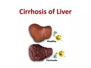
Description • A chronic, progressive disease of the liver • Extensive parenchymal cell degeneration • Destruction of parenchymal cells
Description • Regenerative process is disorganized, resulting in abnormal blood vessel and bile duct relationships from fibrosis
Description • Normal lobular structure distorted by fibrotic connective tissue • Lobules are irregular in size and shape with impaired vascular flow • Insidious, prolonged course
Etiology and Pathophysiology • Cell necrosis occurs • Destroyed liver cells are replaced by scar tissue • Normal architecture becomes nodular
Etiology and Pathophysiology • Four types of cirrhosis: • Alcoholic (Laennec’s) cirrhosis • Postnecrotic cirrhosis • Biliary cirrhosis • Cardiac cirrhosis
Etiology and Pathophysiology • Alcoholic (Laennec’s) Cirrhosis • Associated with alcohol abuse • Preceded by a theoretically reversible fatty infiltration of the liver cells • Widespread scar formation
Etiology and Pathophysiology • Postnecrotic Cirrhosis • Complication of toxic or viral hepatitis • Accounts for 20% of the cases of cirrhosis • Broad bands of scar tissue form within the liver
Etiology and Pathophysiology • Biliary Cirrhosis • Associated with chronic biliary obstruction and infection • Accounts for 15% of all cases of cirrhosis
Etiology and Pathophysiology • Cardiac Cirrhosis • Results from longstanding severe right-sided heart failure
Manifestations of Liver Cirrhosis Fig. 42-5
Clinical ManifestationsEarly Manifestations • Onset usually insidious • GI disturbances: • Anorexia • Dyspepsia • Flatulence • N-V, change in bowel habits
Clinical ManifestationsEarly Manifestations • Abdominal pain • Fever • Lassitude (laziness) • Weight loss • Enlarged liver or spleen
Clinical ManifestationsLate Manifestations • Two causative mechanisms • Hepatocellular failure • Portal hypertension
Clinical ManifestationsJaundice • Occurs because of insufficient conjugation of bilirubin by the liver cells, and local obstruction of biliary ducts by scarring and regenerating tissue
Clinical ManifestationsJaundice • Intermittent jaundice is characteristic of biliary cirrhosis • Late stages of cirrhosis the patient will usually be jaundiced
Clinical ManifestationsSkin • Spider angiomas (telangiectasia, spider nevi) • Palmarerythema
Clinical ManifestationsEndocrine Disturbances • Steroid hormonesof the adrenal cortex (aldosterone), testes, and ovaries are metabolized and inactivated by the normal liver
Clinical ManifestationsEndocrine Disturbances • Alteration in hair distribution • Decreased amount of pubic hair • Axillary and pectoral alopecia
Clinical ManifestationsHematologic Disorders • Bleeding tendencies as a result of decreased production of hepatic clotting factors (II, VII, IX, and X)
Clinical ManifestationsHematologic Disorders • Anemia, leukopenia, and thrombocytopenia are believed to be result of hypersplenism
Clinical ManifestationsPeripheral Neuropathy • Dietary deficiencies of thiamine, folic acid, and vitamin B12
Complications • Portal hypertension and esophageal varices • Peripheral edema and ascites • Hepatic encephalopathy • Fetor hepaticus: is bad breath with a 'dead mouse' or sweet faecal smell. ... It may be caused by severe hepatocellular damage
ComplicationsPortal Hypertension • Characterized by: • Increased venous pressure in portal circulation • Splenomegaly • Esophageal varices • Systemic hypertension
ComplicationsPortal Hypertension • Primary mechanism is the increased resistance to blood flow through the liver
ComplicationsPortal HypertensionSplenomegaly • Back pressure caused by portal hypertension chronic passive congestion as a result of increased pressure in the splenic vein
ComplicationsPortal HypertensionEsophageal Varices • Increased blood flow through the portal system results in dilation and enlargement of the plexus veins of the esophagus and produces varices
ComplicationsPortal HypertensionEsophageal Varices • Varices have fragile vessel walls which bleed easily
ComplicationsPortal HypertensionInternal Hemorrhoids • Occurs because of the dilation of the mesenteric veins and rectal veins
ComplicationsPortal HypertensionCaput Medusae • Collateral circulation involves the superficial veins of the abdominal wall leading to the development of dilated veins around the umbilicus
ComplicationsPeripheral Edema and Ascites • Ascites: - Intraperitoneal accumulation of watery fluid containing small amounts of protein
ComplicationsPeripheral Edema and Ascites • Factors involved in the pathogenesis of ascites: • Hypoalbuminemia • Levels of aldosterone • Portal hypertension
ComplicationsHepatic Encephalopathy • Liver damage causes blood to enter systemic circulation without liver detoxification
ComplicationsHepatic Encephalopathy • Main pathogenic toxin is NH3 although other etiological factors have been identified • Frequently a terminal complication
ComplicationsFetor Hepaticus • Musty, sweetish odor detected on the patient’s breath • From accumulation of digested by-products
Development of Ascites Fig. 42-6
Diagnostic Studies • Liver function tests • Liver biopsy • Liver scan • Liver ultrasound
Diagnostic Studies • Esophagogastroduodenoscopy • Prothrombin time • Testing of stool for occult blood
Collaborative Care • Rest • Avoidance of alcohol and anticoagulants • Management of ascites
Collaborative Care • Prevention and management of esophageal variceal bleeding • Management of encephalopathy
Collaborative CareAscites • High carbohydrate, low protein, low Na+ diet • Diuretics • Paracentesis
Collaborative CareAscites • Peritoneovenous shunt • Provides for continuous reinfusion of ascitic fluid from the abdomen to the vena cava
Peritoneovenous Shunt Fig. 42-8