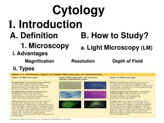
Light Microscopy
Light Microscopy. By: Nicole Sullivan. Modern Light Microscope. Operate with visible light Two magnifying lenses and a variety of correcting lenses High magnification and clarity. Compound Microscope. First lens focuses the image onto the second lens

Light Microscopy
E N D
Presentation Transcript
Light Microscopy By: Nicole Sullivan
Modern Light Microscope • Operate with visible light • Two magnifying lenses and a variety of correcting lenses • High magnification and clarity
Compound Microscope • First lens focuses the image onto the second lens • Second lens magnifies the image and focuses it on the back of the eye • A compound microscope is one that magnifies in stages using several lenses • Note: Compound microscopes can resolve structures that are separated by at least 200 nanometers.
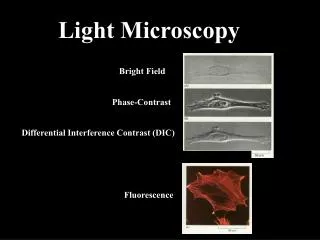
Bright-field microscope Light is transmitted through specimen Little contrast Staining improves contrast, but requires fixed cells, which can alter components
Dark-field microscope • Light is directed at an angle toward specimen • A condenser lens transmits only light reflected off specimen • Field is dark, specimen is light
Phase-contrast microscope Components of microscope bring light waves out of phase. This produces differences in contrast and brightness when light waves recombine
Differential-interference-Contrast microscope Polarized light is split into two beams that have slightly different paths through the sample. Combining these two beams produces greater contrast.
Fluorescence microscope • Fluorescent stains absorb light at one wavelength and emit it at another • Filters transmit only the emitted light
Confocal microscope Light from a laser is focused to a point and scanned across the fluorescently stained specimen in two directions. Produces clear images of one plane of the specimen Other planes are excluded to prevent blurring Multiple planes can be used to reconstruct a 3-D image
Keep in mind… Light microscopes are not powerful enough to resolve many of the structures within cells. This is due to the fact that when two objects are closer than a few hundred nanometers, the light beams reflecting from the two images start to overlap each other.