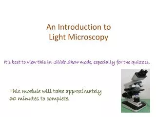
Light Microscopy
Light Microscopy. Uses Before you go to the EM Quick check Maybe all you need Hydrated and living materials. Light Microscopy. Systems Parts Illumination Sample stage Imaging. Light Microscopy. Parts of the light microscope Stand Illuminator Stage Objective(s) Tube Eyepiece(s)

Light Microscopy
E N D
Presentation Transcript
Light Microscopy • Uses • Before you go to the EM • Quick check • Maybe all you need • Hydrated and living materials
Light Microscopy • Systems • Parts • Illumination • Sample stage • Imaging
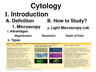
Light Microscopy • Parts of the light microscope • Stand • Illuminator • Stage • Objective(s) • Tube • Eyepiece(s) • Eye, camera
Light Microscopy • Illumination • Through the sample • Incident on surface of sample
Light Microscopy • Through sample illumination • Kohler illumination • Open field and condenser diaphragm • Focus sample • Close field diaphragm and focus with condenser focus control. Use centering controls to center image. • Open field diaphragm to fill image • Take out eyepiece and adjust condenser aperture to ¾ fill BFP of objective
Light Microscopy • Incident Light illumination • Use special objectives • Almost setup for Kohler illumination • Objectives serve as condenser too
Light Microscopy • Objectives • Achromats- RB chromatic and G spherical corrected • Semi-apochromats- RBG chromatic + BG spherical • Apochromats-RBBG chromatic + BG spherical • “Plan” objectives correct for field curvature
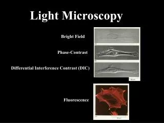
Light Microscopy • Special Techniques • Darkfield (hollow cone illumination)
Light Microscopy • Special Techniques • Phase contrast (blocking phase object interactions with illumination)
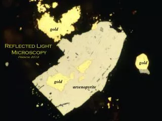
Light Microscopy • Special Techniques • Polarized light (polarized light interaction with birefringent materials)
Light Microscopy • Special Techniques • DIC (Nomarski) (shearing interferometry)