Basic light microscopy
Basic light microscopy. Department of Mechatronics GIST Yong-Gu Lee. References: “ Fundamentals of light microscopy and electronic imaging, ” Chapters 1-6, Douglas B. Murphy, Wiley-Liss, 2001

Basic light microscopy
E N D
Presentation Transcript
Basic light microscopy Department of Mechatronics GIST Yong-Gu Lee • References: • “Fundamentals of light microscopy and electronic imaging,” Chapters 1-6, Douglas B. Murphy, Wiley-Liss, 2001 • “Fundamental of photonics,” Chapter 4 Fourier optics, Bahaa E. A. Saleh and Malvin Carl Teich, John Wiley & Sons, 1991 • http://www.microscopyu.com/ • http://www.bio.unc.edu/courses/2005Spring/Biol188/ • http://support.svi.nl/wiki/ImageFormation • Handbook of optics second edition, McGraw Hill, Vol II, Chapter 17
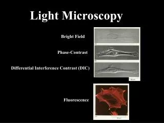
Contents • History • Köhler illumination • Selection of objective lens • Spatial resolution
Hooke microscope had a resolution of about 5 mm “Cells” discovered
perceived and interpretedby the brain as a magnifiedvirtual image about 25 cmin front of the eye
Köhler illumination • The light microscope contains two distinct sets of interlaced focal planes – eight planes in all – between the illuminator and the eyes • Köhler illumination:Illumination that gives bright, uniform illumination of the specimen and simultaneously positions the sets of image and diffraction planes at their proper locations.
Two sets of conjugate focal planes • Four object or field planes: • Viewed by eyepieces (normal, orthoscopic mode) • Four aperture or diffraction planes • Viewed by eyepiece telescope or Bertrand lens, which is focused on the back aperture of the objective lens (aperture, diffraction, conoscopic mode) • The planes are called conjugate, because all of the planes of a given set are seen simultaneously when looking in the microscope
The locations of conjugate focal planes in a light microscope adjusted for Koehler illumination
Köhler illumination • Bright and even illumination in the specimen plane and in the conjugate image plane • Even when illumination is provided by an irregular light source such as a lamp filament, illumination of the object is remarkably uniform across an extended area. Under these conditions of illumination, a given point in the specimen is illuminated by every point in the light source, and, conversely, a given point in the light source illuminates every point in the specimen. • Positioning of two different sets of conjugate focal planes at specific locations along the optic axis of the microscope • This is a strict requirement for maximal spatial resolution and optimal image formation for a variety of optical modes. As we will see, focusing the stage and condenser positions the focal planes correctly, while adjusting the field and condenser diaphragms controls resolution and contrast. Once properly adjusted, it is easier to locate and correct faults such as dirt and bubbles that can degrade optical performance.
Aligning for Köhler illumination You will need an eyepiece telescope or Bertrand lens to examine the lamp filament II: Z(objective) X-Y(condenser) III: Z(condenser) IV: Open(field diaphragm) V: Open/Close (condenser aperture) to set specimen contrast and resolution I : θ–Z-X-Y Ref. http://www.microscopyu.com/tutorials/java/kohler/index.html
Optical Aberrations • Lens errors in modern optical microscopy are an unfortunate problem caused by artifacts arising from the interaction of light with glass lenses. There are two primary causes of non-ideal lens action: Geometrical or Spherical aberrations are related to the spherical nature of the lens and approximations used to obtain the Gaussian lens equation; and Chromatic aberrations, which arise from variations in the refractive indices of the wide range of frequencies found in visible light. Ref: http://micro.magnet.fsu.edu/primer/anatomy/aberrations.html
Coma • In optics (especially telescopes), the coma (aka comatic aberration) in an optical system refers to aberration inherent to certain optical designs or due to imperfection in the lens or other components which results in off-axis point sources such as stars appearing distorted. http://en.wikipedia.org/wiki/Coma_(optics)
Curvature of field • Field curvature indicates that the image plane is not flat, but has the shape of a concave spherical surface as seen from the objective. Different zones of the image can be brought into focus, but the whole image cannot be focused simultaneously on a flat surface as would be required for photography. Field curvature is corrected by the design of the objective and additionally by the tube or relay lens and sometimes the oculars.
Objective correction for optical aberration Ref: http://micro.magnet.fsu.edu/primer/anatomy/objectives.html
Dry objectives must correct for refractions at air/coverslip interface; Oil immersion Increases NA
Airy Disk A simple way to see an “Airy Disk” is to direct a laser beam through a pinhole in a piece of brass--An Airy Disk is the diffraction pattern of a circular hole
Wave optical resolution limits for point sources • Lateral resolution (Rayleigh Limit): • Effect of increasing D/d or decreasing wavelength on resolution of tiny point sources of light Low NA or Long Wavelength ----->High NA or Short Wavelength
Abbe’s theory for image formation in the microscope • Abbe’s Rule: Resolution of a spatial frequency requires interference between the zero and at least one of the first diffraction orders ith order Ref: http://micro.magnet.fsu.edu/primer/java/imageformation/gratingdiffraction/index.html
Summary: wavelength and NA limit resolution in the light microscope • For a fluorescent or self-luminous specimen or for nonluminous points that are examined by bright-field microscopy in transmitted light where the NAcond >= NAobj: -Lateral resolution: p = 0.61l/ NAobj • For a trans-illuminated specimen where the NAcond < NAobj : -Lateral resolution: p = l/ (NAobj + NACond)
Depth of Field and Depth of Focus • When considering resolution in optical microscopy, a majority of the emphasis is placed on point-to-point lateral resolution in the plane perpendicular to the optical axis. Another important aspect to resolution is the axial (or longitudinal) resolving power of an objective, which is measured parallel to the optical axis and is most often referred to as depth of field. • Depth of field is determined by the distance from the nearest object plane in focus to that of the farthest plane also simultaneously in focus. In microscopy depth of field is very short and usually measured in units of microns. The term depth of focus, which refers to image space, is often used interchangeably with depth of field, which refers to object space. Ref. http://www.microscopyu.com/articles/formulas/formulasfielddepth.html
Airy pattern cont’d • The two-dimensional Airy pattern that is formed in the image plane of a point object is, in fact, a cross section of a three-dimensional pattern that extends along the optical axis of the microscope . As one focuses an objective lens for short distances above and below exact focus, the brightness of the central spot periodically oscillates between bright and dark as its absolute intensity also diminishes. Simultaneously, the diameters of the outer rings expand, both events taking place symmetrically above and below the plane of focus in an aberration-free system (Fig . 8) . • Figure 9 c shows an isophot of the longitudinal section of this three-dimensional diffraction image . In the graph we recognize the first minimum of the Airy pattern which we discussed. The intensity distribution along u perpendicular to the focal plane has its first minima at where,
Airy pattern cont’d • The first minimum is at a distance . To transfer distance zi in image space to distance zo in object space, we use the relationship . (Note that for small axial distances, to a close approximation, the axial magnification is the square of the lateral magnification M divided by the refractive index n of the object medium) The distance from the center of the three-dimensional diffraction pattern to the first axial minimum in object space is then given by:
Axial magnification • From imaging equation • Magnification equation Object Image From “Fundamental of photonics,” pp 15, Bahaa E. A. Saleh and Malvin Carl Teich, John Wiley & Sons, 1991
Airy pattern cont’d • The axial resolution of the microscope is closely related to the depth of focus, which is the axial depth on both sides of the image plane within which the image remains acceptably sharp. The depth of focus D is usually defined as ¼ of the axial distance between the first minima above and below focus of the diffraction image of a small pinhole. In the intermediate image plane , this distance is equal to z1 / 2. The depth of focus defined by z1 is the diffraction-limited, or physical, depth of focus .
Geometric depth of focus • It is an inherent property of waves that the image of a point cannot itself be a point, but must be a small circle, the Airy disc. Thus a point cannot be distinguished from a small circle which is called the circle of confusion. Any two points which fall within the radius (not the diameter) of the circle of confusion cannot be resolved as separate points. Di
Depth of focus • These values for the depth of focus, and the distribution of intensities in the three-dimensional diffraction pattern, are calculated for incoherently illuminated (or emitting) point sources (i.e., ) . In general , the depth of focus increases, up to a factor of two, as the coherence of illumination increases (i.e., as ) . • However, the three-dimensional point spread function with partially coherent illumination can depart in complex ways from that so far discussed when the aperture function is not uniform. In a number of phase-based, contrast-generating modes of microscopy , the depth of field may turn out to be unexpectedly shallower than that predicted from above equation and may yield extremely thin optical sections.