Download
1 / 16
160 likes | 345 Vues
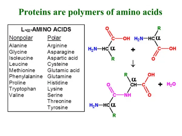
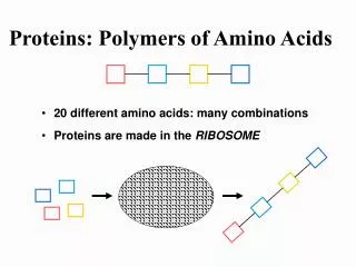
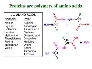
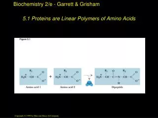
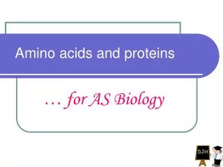
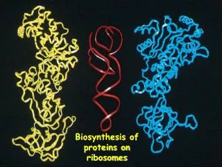
Folding of proteins Proteins are synthesized on ribosomes as linear chains of amino acids. In order to be biologically active, they must fold into a unique three-dimensional structure.

E N D
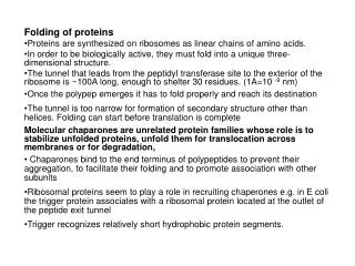
Folding of proteins Proteins are synthesized on ribosomes as linear chains of amino acids. In order to be biologically active, they must fold into a unique three-dimensional structure. The tunnel that leads from the peptidyl transferase site to the exterior of the ribosome is ~100A long, enough to shelter 30 residues. (1A=10 -9 nm) Once the polypep emerges it has to fold properly and reach its destination The tunnel is too narrow for formation of secondary structure other than helices. Folding can start before translation is complete Molecular chaparones are unrelated protein families whose role is to stabilize unfolded proteins, unfold them for translocation across membranes or for degradation, Chaparones bind to the end terminus of polypeptides to prevent their aggregation, to facilitate their folding and to promote association with other subunits Ribosomal proteins seem to play a role in recruiting chaperones e.g. in E coli the trigger protein associates with a ribosomal protein located at the outlet of the peptide exit tunnel Trigger recognizes relatively short hydrophobic protein segments.
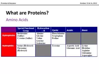
Members of the Heat shock group of proteins are also involved early on , they bind to and protect longer polypeptides. GroEL and GroES facilitate protein folding after translation termination • Chaperones include the heat shock proteins such as HSP-60 and HSP-70 • They stabilize non-native conformation and facilitate correct folding of protein subunits. • They do not interact with native proteins, nor do they form part of the final folded structures. • Some chaperones are non-specific, and interact with a wide variety of polypeptide chains, but others are restricted to specific targets. • They often couple ATP binding/hydrolysis to the folding process. • Essential for viability, their expression is often increased by cellular stress.
GroEl is a large, multi-subunit protein that can accommodate small- and medium-sized proteins • It consists of a central container formed from a stack of rings, and a 'cap' that seals off the folding protein from the external environment • GroEl also has ATP-binding sites that use energy to promote the proper folding and release of the protein
If a protein is trapped in a particularly deep local minimum, we call it ‘misfolded’ as thermal energy may not be enough to kick it out of the state. • There has been tremendous biomedical interest in misfolded proteins as serious human diseases such as Alzheimers’s, Parkinson’s and mad cow disease coincide with the accumulation of misfolded proteins in the form of amyloids. • no consensus on how the amyloids and the disease itself is linked but it is becoming clear that a misfolded protein can be used as a template to cause more misfolded proteins etc, to aggravate the situation. • Theory is that cellular chaperone machineries are overwhelmed by the presence of amyloids so that they do not have enough time to help fold other essential proteins.
There are 3 major classes of chaperones: The Hsp70 familyDnaK is the best known member of this family in E. coli. It is a very highly conserved protein. The Hsp 60 familyThe E. coli member of this family, GroEL forms a double-layered cylindrical structure which provides a protected environment in which proteins can fold. The structure is capped at one end by another heat-shock protein: GroES, a member of the Hsp 10 family. The Hsp 90 family • All are named because these proteins were first identified as Heat Shock Proteins - their synthesis increased in response to a sudden rise in temperature. • In this role, chaperones protect important cell proteins from denaturation as a result of a sudden rise in temperature.
Targeting in Bacteria • As a protein is being synthesized, decisions must be taken about sending it to the correct location in the cell where it will be required. • The information for doing this resides in the nascent protein sequence itself. • Once the protein has reached its final destination, this information may be removed by proteolytic processing. • In bacterial cells, the targeting decision is relatively straightforward: is the protein destined to be an intracellular protein or an extracellular one? • Some more details required in a gram-negative bacterium, such as E. coli, there must be some way of knowing whether a protein is destined to go to the cell membrane, the periplasmic space, or the outer membrane.
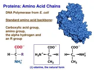
Secreted proteins contain a signal sequence. This is a short (6 - 30) stretch of hydrophobic amino acids, flanked on the N-terminal side by one or more positively charged amino acids and containing neutral amino acids with short side-chains at the cleavage site
As proteins with signal sequences are synthesized, they are bound by the SecB protein. This prevents the protein from folding. SecB delivers the protein to the cell membrane where it is secreted through a pore formed by the SecE and SecY proteins. Secretion is driven by the SecA ATPase. After the protein has been secreted, the signal sequence is removed by a membrane bound leader peptidase.