Functional Imaging in Freely Moving Animals: Advancements and Applications
360 likes | 504 Vues
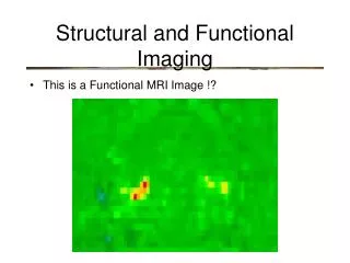
This presentation by R.L. Barbour discusses the innovative techniques in functional imaging for studying freely moving animals. Unlike traditional methods that require restraint, the study emphasizes the importance of capturing a full behavioral repertoire, including aggression, mating, and locomotion, using optical methods. The objectives include determining the feasibility of continuous imaging while simultaneously recording behavioral and neural responses. Utilizing EEG, the research explores the relationships between hemodynamic responses and brain activity, paving the way for dynamic studies of animal behavior and neurophysiology.

Functional Imaging in Freely Moving Animals: Advancements and Applications
E N D
Presentation Transcript
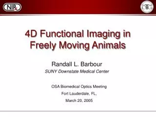
4D Functional Imaging in Freely Moving Animals Randall L. Barbour SUNY Downstate Medical Center OSA Biomedical Optics Meeting Fort Lauderdale, FL, March 20, 2005
Higher Minimal Lower Maximal Levels of Analysis in Biological Investigation Cell Free Preparation Cell Culture Organotypic Culture Degree of Control Phenomenological Complexity Perfused Organ Anesthetized Animal Restrained Animal Freely Moving Animal R.L. Barbour
Why Freely Moving Animals? • Only preparation capable of expressing the full behavioral repertoire of a species. • Aggression • Mating • Fear • Perceptual • Locomotor • Manipulative • Current imaging tools require investigation on restrained/anesthetized animals. • PET/SPECT • MR-fMRI • MEG R.L. Barbour
Why Optical Methods? • Inexpensive, compact instrumentation • High intrinsic sensitivity • Deep tissue penetration • Fast data collection • Easily overlaid on other sensing technologies • Opportunity for dynamic studies R.L. Barbour
Objectives of current study 1. Determine feasibility of continuous functional imaging in freely moving animals while simultaneously recording behavioral, neural and hemodynamic responses. 2. Identify the temporal and spatial dependence of the vascular response as gated to EEG (theta) rhythms. R.L. Barbour
Photo of 9s x 32d imager FRONT Detector channels Timing BACK Power supplies Laser controllers Optical switch Source fiber terminal Lasers / optics R.L. Barbour
Computer w/ frame grabber Laptop computer Figure 12. Schematic of Optical Imaging-EEG-Behavior Monitoring System. DYNOT compact system synchronization Video cam Electro- physiology recording system Optical tether Computer Electrical tether Environmental chamber Head stage w/ Tracking LED Arena w/ animal Schematic of System Setup R.L. Barbour
Optical Fibers 1.8 mm dia. Male part Tracking LED’s Female part Connecting Clips Electrode leads Dual mode optical-EEG measuring head Optical array: 4 source x 16 detector Dual wavelength: 760, 830 nm Framing rate: 17 Hz EEG: 12, 0.1mm diameter electrodes R.L. Barbour
Male Part Optical fiber extension element EEG Electrodes Grounding wires Female Part Dual mode optical-EEG measuring head R.L. Barbour
Olfactory bulbs Right Cortical Hemisphere Receiving Fiber Cerebellum EEG Electrodes Left Cortical Hemisphere Hippocampus Rat Brain Anatomy with Optical-EEG Overlay Transmitting/receiving Fiber R.L. Barbour
Rat with attached helmet and tether R.L. Barbour
Movie of freely moving rat with attached tether R.L. Barbour
Large Irregular Activity Amplitude Time Theta Amplitude Time Hippocampal EEG Rhythms R.L. Barbour
Optical Image Time Series EEG Time Series Time Non-Theta Theta Non-Theta Theta Data Analysis-Integration R.L. Barbour
FEM Mesh for Rat Brain Model S-D Geometry (3D View) FEM Mesh (3D View) 7-compartment model of rat head anatomy obtained from CT scan. 2488 FEM nodes. From Bluestone et al. 2004. R.L. Barbour
Approach • Capture simultaneous: EEG, behavior and dual wavelength tomographic time-series. • Compute volumetric images • Determine temporal/spatial dependence of Hb on EEG/behavior states. R.L. Barbour
RESULTS • Time dependence of spatially integrated findings. • Spatial dependence of temporally integrated findings. R.L. Barbour
Exp. 1: EEG-Gated Hb Spatial Mean Time Series Hboxy Hbdeoxy Hbtot HbO2 Sat Red – Non-Theta Green – Theta (animal moving) R.L. Barbour
Exp 1: Time Averaged-Whole Brain EEG-Gated Hemoglobin Response R.L. Barbour
P-value HbOxy HbDeoxy HbTotal HbSat Stationarity of EEG-Gated Hb Response .. …… R.L. Barbour
Figure 8. Hb response as a function of removal of fraction of initial period. Time Lag of Hb Response R.L. Barbour
Spatially Integrated findings of vascular response to theta rhythm • Increased Hboxy • Decreased Hbdeoxy • Increase Hbtot • Increased HbO2Sat • i.e., BOLD effect R.L. Barbour
EEG-Gated Hb Response Rat 1 Session 1 (Sec 1 - 4) B A Rat 1 Session 2 (Sec 1 - 4) HbOxy HbDeoxy HbTot HbSat Rat 2 Session 2 (Sec 1 - 4) Rat 2 Session 1 (Sec 1 - 4) C D HbOxy HbDeoxy HbTot HbSat R.L. Barbour
Time Dependence of Gated Response Four Sessions Combined (Sec 1 - 4) Four sessions combined (0-1 sec) HbOxy HbDeoxy HbTot HbSat R.L. Barbour
Spatial dependence • Spatial response is reproducible across trials. • Positive, negative and mixed BOLD effects are mainly spatially distinct. R.L. Barbour
Autoregulatory dependent hemoglobin states R.L. Barbour
Spatial Mean Time Series for Autoregulatory State 4 (Balanced) Pixel No Hboxy+ Hbdeoxy+ Hbtot+ R.L. Barbour
Spatial Mean Time Series for Autoregulatory State 5 (Uncompensated oxygen excess) Pixel No Hboxy+ Hbdeoxy- Hbtot+ R.L. Barbour
Spatial Mean Time Series for Autoregulatory State 6 (Compensated oxygen excess) Pixel No Hboxy+ Hbdeoxy- Hbtot- R.L. Barbour
Spatial dependence of autoregulatory response 4 5 Nose 6 3 1 2 R.L. Barbour
Temporal Averaged Gated Maps of Hb States R.L. Barbour
P-values for Theta vs. Non-theta for Autoregulatory dependent hemoglobin states R.L. Barbour
Time-integrated Hb states: Theta 4 5 3 6 Composite 1 2 R.L. Barbour
Time-integrated Hb states: Non-Theta 4 5 3 6 Composite 1 2 R.L. Barbour
Conclusions • Real-time recording of hemodynamic, EEG and behavorial responses is technically feasible in freely moving animals. • Hemodynamic response to theta rhythms are reproducible and spatially distinct. • Method provides for assessment of temporal-spatial dynamics of autoregulatory response to neural activation. R.L. Barbour
Future Considerations • Imaging under defined behavioral paradigms to ascertain localizability of EEG dependent hemodynamic responses. • Influence of pharmacoactive agents on measured responses. • Technological improvements: >S-D pairs, wavelengths, etc. • Development of human compatible system. R.L. Barbour