Download
1 / 40
400 likes | 590 Vues
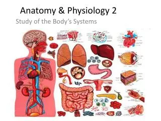
Anatomy and Physiology For The First Class 2 nd Semester. Urinary System. Urinary System. It is one main excretory system in the body. It consists of: 2 kidneys which secret urine. 2 ureters , which convey the urine from the kidney s to the urinary bladder.

E N D
Anatomy and Physiology For The First Class 2nd Semester
Urinary System • It is one main excretory system in the body. • It consists of: • 2 kidneys which secret urine. • 2 ureters, which convey the urine from the kidney s to the urinary bladder. • The urinary bladder where urine collects and is temporarily stored. • The urethra through which the urine passes from the urinary bladder to the exterior.
The Main Functions of the Urinary System Most functions of the urinary system are produced by kidneys • Excretion: removal of waste products (including nitrogenous compounds, urea uric acid, excess ions and some drugs) from body fluids. • Homeostasis of water and electrolyte concentrations within the body by: • regulation of blood volume and pressure. • regulation of plasma ion concentration. • Stabilizing blood pH. • Conserving nutrients. 3. Production of hormones: Renin, erythropoietin, thrombopoietin 4.Activate vitamin D.
kidneys • Kidneys are bean-shaped organs, about 11 cm long, 6 cm wide, 3 cm thick and weight 150 g. in the medial border of each kidney there is a renal helium. • located just above the waist between the peritoneum and the posterior wall of the abdomen • Because their position is posterior to the peritoneum of the abdominal cavity they are said to be retroperitoneal organs. • The kidneys are located between the level of the last thoracic and third lumbar vertebrae • The right kidney is slightly lower than the left. • Each kidney is surrounded by a renal capsule, adipose tissue and renal fascia. • The kidneys lie on either side of the vertebral column, so each kidney is associated with a different group of structures: Right kidney: superiorly – the right adrenal gland Anteriorly – the right lobe of liver, the duodenum and the hepatic flexure of the colon. Posteriorly – the diaphragm, and the muscles of the posterior abdominal wall. Left Kidney: Superior – the left adrenal gland Anteriorly –the spleen, stomach, pancreas, jejunum and splenic flexure of the colon. Posteriorly - the diaphragm and muscles of the posterior abdominal wall.
Internal Anatomy of the kidneys • In longitudinal section of the kidney appears two regions: • Cortex(light red region) 2. Medulla (dark red brown region): there are several pyramids in the renal medulla. The apex of each pyramids is called renal papilla, points toward renal hilum. • Renal papillae are surrounded by cup like structures called minorcaylces. • Each kidney has 8-18 minor calyces which combine together to form 2-3 major calyces. • The major calyces combine to form in a single large cavity called renal pelvis. • From the renal pelvis the ureter is originated.
Blood and Nerve Supply • The kidneys receive 20-25% of resting cardiac out put via right and left renal arteries. • In the adult the blood flow through the kidneys is about 1200 ml/ min. • Renal nerves are part of sympathetic division of autonomic nervous system.
Microscopic structure of the kidneys • The functional unit of the kidney is called nephron. • There are 1 – 2 million nephrons in each kidney. • The collecting ducts transport urine through pyramids to the calyces and renal pelvis.
The Nephron The nephron consists of: 1. renal corpuscle (Malpighian corpuscle) 2. renal tubule
Blood inter glomeruli through afferent arterioles and circulate through glomerular capillaries than the blood leave glomeruli through efferent arterioles. • In the afferent arterioles there are specialized cells called juxtaglomerular cells (release renin). • Juxtaglomerular cells are part of juxtaglomerular appratus. The renal corpuscle consists of: a). Tuft of blood capillaries called the glomerulus. b). A cup like, double layered covering for the glomerulus called the glomerular capsule (Bowman’s capsule)
The renal tubule is divided into several parts: Proximal convoluted tubule (PCT) Loop of Henle: descending limb, thin ascending limb and thick ascending limb Distal convoluted tubule (DCT): the portion of DCT near juxtagolmerular cells contains special area called macula densa. macula densa cells are sensitive to the concentration of sodium chloride in the DCT The juxtaglomerular cells and macula densa of DCT is formed the juxtaglomerular apparatus.
Functions of the kidneys 1. Urine formation The composition of urine reflect exchange of substances between the nephron and the blood in the renal capillaries. Waste products of protein metabolism are excreted, electrolyte levels are controlled and pH (acid base balance) is maintained by excretion of H ions. to produce urine, there are three basic processes: • Glomerular filtration • Tubular reabsorption • Tubular secretion • Urine excretion
Glomerular filtration • Glomerular filtration occurs in the semipermeable walls of the glomerulus and glomerular capsule. • Water and other small molecules (salts, ions, glucose, amino acids, creatinin, urea, and others) pass through these walls, while blood cells, plasma proteins and other large molecules are too large to filtrate. • Filtration takes place because there is difference between blood pressure in the glomerulus and the pressure of the filtrate in the glomerular space (urinary space). • Three pressures in the renal corpuscle control the filtration process these are: Blood hydrostatic pressure = 55 mmHg Blood osmotic pressure = 30 mmHg Capsular (filtrate) hydrostatic pressure = 15 mmHg • The net filtration pressure (NFP) is: NFP = BHP – ( BOP + FHP) = 55- (30 + 15) = 10 mmHg
The volume of filtrate formed by kidneys each minute is called Glomerular Filtration Rate (GFR). • Normally GFR = 125 ml/ minute i.e. 180 liters/ day of filtrate by two kidneys. • Factors influencing GFR: 1. Hydrostatic pressure 2. Osmotic pressure gradient 3. Permeability of capillaries GFR is increased during pregnancy as metabolites produced in the fetus have also to be excreted. GFR is decreased in hypotension and in advanced renal diseases. • Nearly all the filtrate is later reabsorbed and only less 1% i.e. 1.5 liters excreted as urine.
Tubular Reabsorption and Tubular Secretion • Tubular reabsorption: means selectively moving substances like nutrients, electrolytes and others from filtrate (nephron tubule) back into blood. This process is important for maintenance the consistency of internal environment of the body. • Tubular secretion: means selectively moving substances from blood to the filtrate (nephron tubule). • in the proximal convoluted tubule: reabsorption: K ion, H2O (65%), NaCl (65%), HCO3 ions, amino acids (100%), glucose (100%). secretion: Uric acid, organic acids. The majority of reabsorption occurs in PCT • In the Loop of Henli: reabsorption:H2O (descending part of Henli) and NaCl 25% (ascending part of Henli) • In the distal convoluted tubule (DCT): reabsorption :NaCl (5%), H2O Secretion :K ion, H ion. the majority of secretions occurs in DCT. • Collecting tubules: reabsorption:small amaunts of Urea, and NaCl (5%), and H2O.
Composition of urine • Urine is clear and amber in color (due to presence of urobilin). The pH is around 6 (normal range of 4.5 – 8). • Urine production is decreased during sleeping and exercise. • The components of urine are: Water 96% Urea 2% (Uric acid, Creatinine , Ammonia , Sodium , Potassium, Chlorides , Phosphates , Sulphates, Oxalates) 2%.
Hormones that Influence Selective Reabsorption • Parathyroid hormone and Calcitonin: these hormones regulate the reabsorption of calicium and phoshate from distal collecting tubules. • Antidiuretic Hormone (ADH): • secreted by posterior lobe of pituitary gland. • acts on distal convoluted tubules and collecting tubules that leads to increase the reabsporption of water. • secretions of ADH is controlled by a negative feed back mechanism. • Aldosterone: • Secreted by adrenal cortex • Increases the reabsorption of sodium and water and the excretion of potassium. • Secretion is regulated through negative feed back mechanism. • Atrial natriuretic peptide (ANP) • Secreted by the atria of the heart in response to increase blood volume. • Acts to decrease reabsorption of sodium and water from the proximal convoluted tubules and collecting tubules. • Secretion of ANP is also regulated by negative feedback mechanism.
2. Maintenance of Water Balance : Decrease water in the body leads to: 1. increase blood osmolarity over 300 mOsm/ L. 2. osmotoreceptor in the hypothalamus are sensitive to changes in the blood osmolarity so two things occur: feeling thirst and secretion of ADH. 3. ADH enhances water reabsorption by renal tubules. Therefore the volume of urine becomes decrease
When blood volume is increased stretch receptors in the atria of the heart release atrial natriuretic hormone (ANP). ANP reducesreabsorption of sodium and water by the proximal convoluted tubules and collecting ducts (i.e. more sodium and water are excreted).
3. Control of Blood Pressure • Macula densa cells of juxtaglomerular apparatus are very sensitive to decreasein sodium and in decrease in blood pressure. • When blood pressure is low the cells of macula densa send stimulus to the juxtaglomerular cells to secret renin. • Renin acts on protein (synthesized by the liver) is called angiotensinogen. • Renin converts angiotensinogen to the angiotensin I, then angiotensin I converted to angiotensin II by enzyme called angiotensin converted enzyme (produced by lungs). • angiotensin II acts by two ways to raised blood pressure 1. directly on the blood vessels and leads to vasoconstriction. 2. enhances adrenal cortex to release aldosterone hormone. • Aldosterone acts on renal tubules to increase reabsorption of sodium and water.
4. Acid - Base Balance • Under normal conditions the pH of urine is acidic. • The kidneys regulate the blood pH by three main mechanisms: • Reabsorption of the filtered HCO3 ions. • Generation of NaHCO3 ions • Excretion of acid as H ions as titrable acid and as NH4 ammonium. • The kidneys regulate blood pH by increasing or decreasing the excretion of hydrogen and bicarbonate as required. If the pH falls hydrogen ion excretion is increased and bicarbonate conserved. 5. other mains functions of the kidneys is secreted hormones and activated vitamin D3.
Urine transportation, Storage and Elimination • Urine drains through papillary ducts into the minor calyces, which join to become major calyces that unite to form the renal pelvis. • From the renal pelvis urine first drains into the ureters and then into urinary bladder. • Then urine is discharged from the body through the single urethra.
Ureters • The ureters (right and left) are long tubes which convey the urine from the renal pelvis to urinary bladder. • Each ureter is about 25 -30 cm long and about 4-5 mm in diameter. • Parts of ureter: ₋ abdominal part extends from the renal pelvis to the brim of bony pelvis ₋ pelvis part at the brim of pelvis the ureter crosses the upper end of the external iliac artery and vein and comes to the lie on the lateral wall of the pelvis than runs forwards to reach the urinary bladder. ₋ intracystic part is the small of the ureter which passes obliquely through the posterior wall of urinary bladder. • There are no anatomical valve at the opening of each ureter into the the urinary bladder. • obliquely passage of the intracystic part of ureter leads to form a physiological valve which closes the ureter opening when the bladder filled with urine and prevents the backflow of urine. • Ureter wall consists of three coats: outer fibrous coat middle muscular coat inner mucous coat • Peristaltic contractions of the muscular layer of the ureter lead to urine transportation to the bladder.
Urinary Bladder • The urinary bladder is a reservoir for urine, it lies in the pelvic cavity in front of rectum in male and uterus in female. Its size and position vary, depending of the volume of the urine it contains, when distended the bladder rises into the abdominal cavity. • The peritoneum covers only the superior surface of the bladder. • The bladder is roughly bear shaped but becomes more oval as it fills with urine. • Inner surface of the bladder is arranged in folds when the bladder is empty and gradually disappear as it fills. • The total capacity of the bladder is about 600 ml • The bladder opens into the urethra at its lowest point (the neck). • The three orifice in the bladder wall form a triangle or trigone. The upper two orifices on the posterior wall ore the opening of the ureters and the lower orifice is the opening in to the urethra. • The bladder wall is composed three layers: • 1. the outer layer of loose connective tissue containing blood and lymphatic vessels and nerves. • 2. the middle layer is formed by smooth muscle fibers which are arranged in the three layers , the fiber of external internal fibers are arranged longitudinally while the middle fibers arranged circularly. Contraction of this muscle caot is responsible for empty of the bladder. • 3. mucosa (transitional mucous membrane).
Urethra and its Sphincters • The tube that connect the lower end (neck) of the bladder to the exterior. • Male urethra is about 20 cm in length, is divided into 3 parts: prostatic urethra (3 cm), membranous urethra (1.25 cm), and penile urethra (15.75 cm). Membranous urethra is surrounded by external sphincter. • The male urethra is associated with the urinary and the reproductive system • Female urethra is about 3.8 cm long. It extends from the neck of the bladder to the external meatus. It transverses the external sphincter and lies immediately in front of the vagina. • Internal sphincter of the urethra : it is formed from thickening in the circular smooth muscle in the area of the neck of the bladder. • External sphincter of the urethra: it is formed from skeletal muscle fibers (voluntary). This sphincter provides voluntary control over micturition.
Micturition • when 300 -400 ml of urine have accumulated in the bladder, sensory nerve fibers in the bladder wall sensitive to stretch are stimulated. • In the infant this initiates spinal reflex and micturation occurs, urine passed in the response to the parasympathetic stimulation of the bladder causing contraction of the detrusor muscle and relaxation of the internal sphincter, urine is expelled from the bladder and passes through the urethra before leaving the body. • When the nervous system is fully developed the micturition reflex is stimulated but sensory impulses also pass upwards to the brain and there is awarness of the need to pass urine. By learned and conscious effect contraction of the external urethral sphincter and muscles of the pelvic floor can inhibit micturition until it is convenient to empty the bladder. When detrusor muscle contract and there is reflex relaxation of the internal sphincter and voluntary relaxation of the external sphincter.