Download
1 / 57
580 likes | 685 Vues
This overview delves into the essential components of the human circulatory system, focusing on the heart, blood vessels, and lymphatic vessels. The circulatory system performs critical functions including transporting oxygen and nutrients to cells, removing waste products, and distributing hormones. We explore the heart's anatomy, comprising the myocardium and endocardium, and the mechanisms of heart contractions. Additionally, we examine the conduction of electrical impulses, the role of valves, and calculate cardiac output to highlight the system’s efficiency and responsiveness during rest and exercise.

E N D
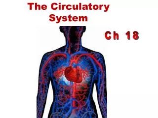
The Circulatory System Ch 18
Components of the Human Circulatory System The Heart Blood Vessels Blood Lymphatic Vessels Lymph
Functions of the Circulatory System • Transport oxygen to cells • Transport nutrients from the digestive system to body cells • Transport hormones to body cells • Transport waste from body cells to excretory organs • Distribute body heat
Energy Requirements: • lots of mitochondria • aerobic respiration
Mechanisms and events of contractions: • All or none law- at organ level, not cellular level • Means of stimulation- autorhythmicy • Length of refractory period- cardiac (250ms), skeletal (1-2ms)
Location of Heart Location of Heart
Layers of Cardiac Tissue • Visceral pericardium • Outer protective layer composed of a serous membrane • Includes blood capillaries, lymph capillaries, and nerve fibers.
Layers of Cardiac Tissue • Myocardium • Relatively thick. • Consists largely of cardiac muscle tissue responsible for forcing blood out of the heart chambers. • Muscle fibers are arranged in planes, separated by connective tissues that are richly supplied with blood capillaries, and nerve fibers.
Layers of Cardiac Tissue • Endocardium • Consists of epithelial and connective tissue that contains many elastic and collagenous fibers. • Connective tissue also contains blood vessels and some specialized cardiac muscle fibers called Purkinje fibers. • Lines all of the heart chambers and covers heart valves.
Heart Anatomy http://www.youtube.com/watch?v=tBQa8IBzP6I
Heart Anatomy Left ventricle Right ventricle Interventricular septum
Mechanisms & Events of Contraction • Means of stimulation • Organ vs motor unit contraction • Length of absolute refractory period
Microscopic Anatomy of Cardiac Muscle • Cardiac muscle cells are striated, short, fat, branched, and interconnected • Connective tissue matrix (endomysium) connects to the fibrous skeleton • T tubules are wide but less numerous; SR is simpler than in skeletal muscle • Numerous large mitochondria (25–35% of cell volume)
Nucleus Intercalated discs Cardiac muscle cell Gap junctions Desmosomes (a) Figure 18.11a
Microscopic Anatomy of Cardiac Muscle • Intercalated discs: junctions between cells anchor cardiac cells • Desmosomes prevent cells from separating during contraction • Gap junctions allow ions to pass; electrically couple adjacent cells • Heart muscle behaves as a functional syncytium
VALVES Pulmonary valve Myocardium Aortic valve Tricuspid (right atrioventricular) valve Area of cutaway Mitral valve Tricuspid valve Mitral (left atrioventricular) valve Myocardium Tricuspid (right atrioventricular) valve Aortic valve Mitral (left atrioventricular) valve Pulmonary valve Aortic valve Pulmonary valve Aortic valve Pulmonary valve Area of cutaway (b) Fibrous skeleton Mitral valve Tricuspid valve (a) Anterior Figure 18.8a
VALVES Pulmonary valve Aortic valve Area of cutaway Mitral valve Tricuspid valve Chordae tendineae attached to tricuspid valve flap Papillary muscle (c) Figure 18.8c
pulmonary arteries right atrium aorta pulmonary arteries pulmonary vein superior vena cava left atrium left ventricle inferior vena cava right ventricle
Heart Valves pulmonary semilunar valve aortic semilunar valve bicuspid valve tricuspid valve
Cardiac Output CO = the vol. of blood ejected from the l. or r. ventricle into the aorta or pulmonary trunk each min. CO= SV x HR SV= stroke vol.; the vol of blood ejected from the ventricle during each contraction (ml/beat) HR= heart rate; # beats/min, at rest ~60, exercise ~100
Cardiac Output (at rest) SV = 75 ml/beat HR = 75 beats/min CO = 75 ml/b x 75 b/min CO = 5250 ml/min = 5.25 L/min
Cardiac Output (exercise) SV = 100 ml/beat HR = 100 beats/min CO = 100 ml/b x 100 b/min CO = 10 L/min
Nerve Innervation: pons Vagus nerve from medulla (parasympathetic division) acetylcholine (slows heart) Cardioacceleratory center in medulla (sympathetic) adrenaline from adrenal glands (speeds up heart) medulla oblongata vagus
Electrocardiogram (ECG) 0.1 sec 0.3 sec 0.4 sec • P = atrial depolarization ~ 0.1 sec atria contracts • QRS = ventricular depolarization ventricles contract (lub), contraction stimulated by Ca++ uptake • T = ventricular repolarization ventricles relax (dub)
Excitation of the Heart R Depolarization SA node Repolarization T P Q S 1 Atrial depolarization, initiated bythe SA node, causes the P wave. Figure 18.17, step 1
R Depolarization SA node Repolarization T P Q S 1 Atrial depolarization, initiated bythe SA node, causes the P wave. R AV node T P Q S 2 With atrial depolarization complete,the impulse is delayed at the AV node. Figure 18.17, step 2
R Depolarization SA node Repolarization T P Q S 1 Atrial depolarization, initiated bythe SA node, causes the P wave. R AV node T P Q S 2 With atrial depolarization complete,the impulse is delayed at the AV node. R T P Q S 3 Ventricular depolarization beginsat apex, causing the QRS complex.Atrial repolarization occurs. Figure 18.17, step 3
Depolarization Repolarization R T P Q S 4 Ventricular depolarization iscomplete. Figure 18.17, step 4
Depolarization Repolarization R T P Q S 4 Ventricular depolarization iscomplete. R T P Q S 5 Ventricular repolarization beginsat apex, causing the T wave. Figure 18.17, step 5
Depolarization Repolarization R T P Q S 4 Ventricular depolarization iscomplete. R T P Q S 5 Ventricular repolarization beginsat apex, causing the T wave. R T P Q S 6 Ventricular repolarization iscomplete. Figure 18.17, step 6
(a) Normal sinus rhythm. (b) Junctional rhythm. The SA node is nonfunctional, P waves are absent, and heart is paced by the AV node at 40 - 60 beats/min. (d) Ventricular fibrillation. These chaotic, grossly irregular ECG deflections are seen in acute heart attack and electrical shock. (c) Second-degree heart block. Some P waves are not conducted through the AV node; hence more P than QRS waves are seen. In this tracing, the ratio of P waves to QRS waves is mostly 2:1. Figure 18.18
Heart Sounds • Two sounds (lub-dup) associated with closing of heart valves • First sound occurs as AV valves close and signifies beginning of systole • Second sound occurs when SL valves close at the beginning of ventricular diastole • Heart murmurs: abnormal heart sounds most often indicative of valve problems
Coronary Artery Disease(CAD) • Arteriosclerosis • HDL vs LDL
Homeostatic Imbalances • Angina pectoris • Thoracic pain caused by a fleeting deficiency in blood delivery to the myocardium • Cells are weakened • Myocardial infarction (heart attack) • Prolonged coronary blockage • Areas of cell death are repaired with noncontractile scar tissue http://www.youtube.com/watch?v=l36zIKP53Ls
Coronary Artery Disease(CAD) Diagnosis • Stress test • Echocardiography • http://youtu.be/DuanD-z45tw • Cardiac catheterization • Coronary angiography
Coronary Artery Disease(CAD) Treatment • Coronary bypass grafting (CABG) • Percutaneous transluminal coronary angioplasty (PTCA)


