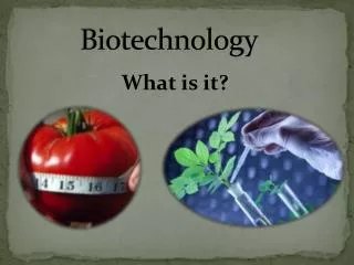
Biotechnology
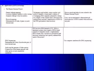
This lecture delves into the crucial steps of genetic engineering, focusing on how to isolate a desired gene, such as human insulin, and utilize plasmid vectors for its insertion into E. coli. We will explore methods like restriction enzyme cleavage and gel electrophoresis to achieve gene insertion, followed by transformation and testing of engineered microbes. Emphasizing the significance of these biotechnological tools, this discussion highlights the continuous advancements in genetic engineering that enhance our ability to produce essential proteins and enzymes for medicinal and industrial use.

Biotechnology
E N D
Presentation Transcript
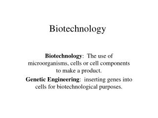
Biotechnology Biotechnology: The use of microorganisms, cells or cell components to make a product. Genetic Engineering: inserting genes into cells for biotechnological purposes.
Introduction • Today’s lecture will focus on Fig. 10.9 on page 297. • We will talk about how we get from isolating a desired gene product to the actual production of the protein or enzyme. • Realize though, that the techniques we talk about as well as the others mentioned in your textbook are only a few of the tools available for research and genetic engineering, and others are being developed all the time.
Overview of Genetic Engineering • Let’s say we want to isolate the gene for human insulin and put it into E.coli so it can make insulin. The following are the basic steps required to do that. • 1. Select a vector (plasmid) for our desired gene. • A vector is a piece of DNA that acts like a taxi for a selected gene. • 2. Original DNA cleaved by restriction enzymes to isolate the desired gene. • 3. The chosen vector is cleaved by the same restriction enzymes and the selected gene is inserted. • 4. The vector is transformed into the target cell. • 5. Test the microbe for presence of the new gene. • 6. The engineered microbe is grown and gene copies are harvested OR microbe makes protein product and the protein product is harvested.
1. Isolate target vector • Plasmid vectors can be obtained from microbes themselves, or by ordering them commercially. • If a the vector is to be obtained from a microbe, that microbe is grown in mass. • The the microbes are lysed, releasing their intracellular contents. • Then the plasmid is isolated from protein and membrane debris
If the plasmid is obtain commercially it means that it was previously grown in a microbe and isolated. • The advantage to buying a plasmid commercially is that it’s nucleotide sequence is already known. This is called a map. See Fig. 10.8 for an example. • The map shows what genes are on the plasmid, the size, as well as the sites called restriction enzyme sites. • The restriction enzyme sites are very important for gene insertion into the plasmid.
2. Original DNA cleaved by restriction enzymes • The next step is to isolate the selected gene that we want to put into E.coli. So if we use the insulin example, we want to isolate the gene for insulin. • Restriction enzymes are designed to cut DNA at a specific sequence. • Restriction enzymes are essentially DNA scissors. • Restriction enzyme sites are like the little diagram of scissors on a paper project that indicate that you are to cut on the dotted line.
Each restriction enzyme will only cut at the DNA sequence it recognizes. See Fig. 10.1. • ON the plasmid map you will see many restriction enzymes marked. Some examples are: • EcoRI and RII: restriction enzymes made by E.coli; • HaeI, II, III: restriction enzymes made by Haemophilus influenzae; • SalI: a restriction enzyme made by Salmonella; • The insulin gene and it’s surrounding genetic sequence is analyzed to determine which restriction enzyme sites surround the gene and to cut it out.
Gel Electrophoresis • After the insulin gene is cut out, it is still surrounded by cut DNA and restriction enzymes. It needs to be isolated so that it can be inserted into the plasmid vector. • Gel Electrophoresis is used to separate selected genes from the rest of the cell garbage.
The gel that is used for Gel Electrophoresis is made up of a molecule called agarose. • To help you understand what is happening better let’s talk about “Jello Jigglers”. • For those of you that don’t know what Jello Jigglers are, they are shapes made from concentrated jello. • In other words, less water is added, meaning that the sugar and gelatin molecules are more concentrated giving a stiffer jello. The stiffer jello allows you to cut shapes out of the jello.
An agarose gel is similar but instead of gelatin and sugar, agarose is used to create the “jello”. The more water that is added, the less concentrated the agarose is. • Just like when you make jello, the agarose and water are heated up first. Then they are poured into a mold to cool. The molds come in various sizes but essentially they are all rectangles that are about 1 ½ inches thick.
Once the agarose is poured into the mold a “comb” is put into the agarose at one end of the gel. It looks similar to a hair comb but there are usually only 8 or 10 teeth that are very wide and flat instead of pointed at the ends. • When the agarose is set, the comb is removed and small slits remain where the teeth were. These slits are called wells and are the place where the DNA is put into the gel. • A “ladder” is put into one of the wells. The “ladder” is commercially made by putting DNA fragements of various known sizes together. These DNA fragments help the researcher to know which piece of DNA in the other wells is the DNA with the insulin gene. (We know the size of the gene because we have its sequence.)
Ok. So we load the ladder in one well and the rest of the wells we fill with the DNA and garbage. • Each DNA nucleotide has a phophate group on it. Remember from the cell membrane that a phophate group has a slight negative charge. So the charge on DNA is negative. • We cover the gel with a buffer that conducts electricity and then put a negative charge on the side of the rectangle closest to the DNA in the wells and a positive charge on the opposite end of the rectangle. • This arrangement causes the DNA to move down the gel away from the negative charge, towards the positive charge.
The more agarose in the gel, the more molecules there are that the DNA has to move through. • The smaller pieces of DNA can move through much more quickly than the large pieces of DNA. • The large DNA remains closer to the wells and the small DNA moves closer to the opposite end. • After the DNA has been separated on the gel it is stained so that we can see where the DNA is. See Fig. 10.2.
By using the ladder and comparing the known sizes of DNA on the ladder to the known size of the insulin gene, we can find the insulin gene. • Once the insulin gene is identified on the gel it can be excised out of the gel by using a razorblade. Then the agarose is melted and the DNA removed and cleaned.
Polymerase Chain Reaction (PCR) • The last step before the insulin gene can be inserted into the plasmid vector is PCR. • PCR is a quick and easy way to make multiple gene copies (DNA replication). See Fig. 10.6. • 1. First you take a small plastic tube and put in the insulin gene. It will be the template. • 2. Then you add some primers. Remember in DNA replication that a primer is a hand for the other nucleotides to hold onto so that they can start replication of the template. The primers that we use for our gene are specially made so that they recognize and bind to the template gene sequence at the 5’ end.
3. Then some DNA bases are put into the tube. (ATCG) DNA can’t be made without them! • 4. Then a polymerase is added. Just like in DNA replication, the polymerase puts the nucleotides together. • Now were ready to begin the process of PCR. • The first step is to heat the DNA. • The heat breaks the DNA strands apart.
Then the sample is cooled and the primers bind to the template strands. Then the polymerase begins to attach the nucleotides to make complementary strands. • Then the cycle begins again. The sample is heated up, the strands separate. The sample is cooled and the primers and polymerase synthesize complementary strands. This process is repeated many times to create many gene copies. • PCR used to be done using water baths heated to the necessary temperatures. Then a timer would be set for a couple of minutes and the researcher would have to move the sample from bath to bath cycle after cycle. Now days there is a special machine made just for cycling the temperatures for PCR
After the PCR is finished, another round of gel electrophoresis is performed to get rid of the enzymes and unused nucleotides. • Now the gene is read to be inserted into the plasmid vector. • It’s the moment you have been waiting for and you get to wait some more until next lecture. I hope you’re not too disappointed. • Homework will be given after the next lecture.