Download
1 / 26
270 likes | 633 Vues
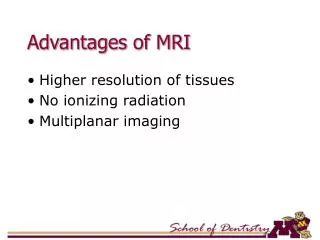
Advantages of MRI. Higher resolution of tissues No ionizing radiation Multiplanar imaging. Disadvantages of MRI. Long imaging time Hazards with ferromagnetic metals (pacemakers, vascular clips, etc) Claustrophobia Higher cost. Relative Brightness of Tissues. Fat White Marrow Brain

E N D
Advantages of MRI • Higher resolution of tissues • No ionizing radiation • Multiplanar imaging
Disadvantages of MRI • Long imaging time • Hazards with ferromagnetic metals (pacemakers, vascular clips, etc) • Claustrophobia • Higher cost
Relative Brightness of Tissues Fat White Marrow Brain Muscle Gray Body Fluid TMJ Disk Cortical Bone Air Black
Nuclear Medicine • Radioactive compounds • Target tissues • Radioactive agents pools in the target tissues • Detected and imaged by external detectors (gamma camera).
Nuclear Medicine • Shows structure and function of the target tissues • Static and dynamic conditions • Scintigraphy scans or RN (radionuclide) scans • Bone scans or salivary gland scans
Technetium • 99mTcO4- - thyroid and salivary gland scan • 99Tc phosphate - bone scan
Phases of Salivary Gland Scan • Flow phase: • Five to 10 mCi of 99mTcO4 • first 30 to 120 seconds • shows flow of blood • Concentration phase: • next 30 to 45 minutes • demonstrate the anatomy and function • Washout Phase: • administer sialagogue • demonstrates secretory capabilities
Cephalometric Radiography • Reproducible and standardized views • For measurements and assess growth • Fixed source to film distance – 60 inches • Cephalostats and earplugs help in reproducible positions
Contrast Agents • Radiopaque materials • Water soluble • Fat soluble • 28 – 38% iodine
Phases of Sialography • Ductal • Acinar • Evacuation
Indications of Sialography • Acute swelling secondary to ductal obstruction • Recurrent Inflammation • Palpable salivary gland mass • Autoimmune Sialadenitis
Contraindications of Sialography • Sensitivity to contrast agents • Acute Sialadenitis • Limited use in tumor diagnosis
Arthrography • Contrast media is introduced in joint spaces • Upper vs. lower joint space • Viewed by Image Intensifier Fluoroscopy • Video recording allows study of joint movement
Open Position • Translation of condyle • Reduction of disk