Necrosis and apoptosis
(Foundation Block, pathology). Lecturer name: Dr. Maha Arafah Lecture Date: 17-9-2012. Necrosis and apoptosis. Cell injury L2. Objectives. List causes of cell injury List mechanisms of cell injury Understand the changes in reversible and irreversible cell injury

Necrosis and apoptosis
E N D
Presentation Transcript
(Foundation Block, pathology) Lecturer name: Dr. MahaArafah Lecture Date: 17-9-2012 Necrosis and apoptosis Cell injury L2
Objectives • List causes of cell injury • List mechanisms of cell injury • Understand the changes in reversible and irreversible cell injury • Define necrosis and apoptosis • List the different conditions associated with apoptosis, its morphology and its mechanism • List the different types of necrosis, examples of each and its features • Know the difference between apoptosis and necrosis
Etiologic agents DEFICIENCY OF OXYGEN Ischemia vs. Hypoxia PHYSICAL AGENTS CHEMICAL AGENTS INFECTION IMUNOLOGICAL REACTIONS GENETIC DERANGEMENTS NUTRITIONAL IMBALANCE AGING
Brain – massive haemorrhagic focus (ischemia) in the cortex This is a lesion caused by DEFICIENCY OF OXYGEN
Abscess of the brain (bacterial) This is a lesion caused by infectious agent
Hepatic necrosis (patient poisoned by carbon tetrachloride) This is a lesion caused by chemical agent
Pulmonary caseous necrosis (coccidioidomycosis) This is a lesion caused by infectious agent
Gangrenous necrosis of fingers secondary to freezing This is a lesion caused by physical agent
The “boutonnière” (buttonhole) deformity This is a lesion caused by intrinsic factors (autoimmune disease)
Liver: macronudular cirrhosis (HBV) This is a lesion caused by infectious agent: Viral hepatitis (chemical:alcohol, genetic:a1-AT deficiency)
myocadial cells: loss of function after 1-2 min of ischemia However do not die until 20 to 30 min of ischemia EM: 2-3 hours, LM 6-12 hours
Morphologic changes in Reversible Injury Early changes: (1) Cloudy swelling or hydropic changes: Cytoplasmic swelling and vacuolar degeneration due to intracellular accumulation of water and electrolytes secondary to failure of energy-dependent sodium pump. • (2) Mitochondrial and endoplasmic reticulum swelling due to • loss of osmotic regulation. • (3) Clumping of nuclear chromatin.
Vacuolar (hydropic) change in cells lining the proximal tubules of the kidney Reversible changes
Morphologic changes in irreversible injury: 1. Severe vacuolization of the mitochondria, with accumulation of calcium-rich densities. 2. Extensive damage to plasma membranes. 3. Massive calcium influx activate phospholipase, proteases, ATPase and endonucleases with break down of cell component. 4. Leak of proteins, ribonucleic acid and metabolite. 5. Breakdown of lysosomes with autolysis. 6. Nuclear changes: Pyknosis, karyolysis, karyorrhexis.
Morphologic changes in irreversible injury - Dead cell are either collapsed and form a whorled phospholipid masses or degraded into fatty acid with calcification. - Cellular enzymes are released into circula- tion. This provides important clinical parameter of cell death e.g. increased level of creatininkinase in blood after myocardial infarction
Myocardial infarct Cell Pathology This is a lesion caused by oxygen deprivation
Mechanisms of cell injury • Depletion of ATP • Damage to Mitochondria • Influx of Calcium • Free radicals production • Disruption of cell membrane and chromatin
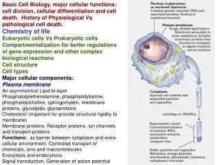
Cell Death • Death of cells occurs in two ways: • Necrosis--changes produced by enzymatic digestion ofcells after irreversible injury • Apoptosis--vital process that helps eliminate unwanted cells--an internally programmed series of events effected by dedicated gene products Autolysis Autolysis is the death of individual cells and tissues after the death of the whole organism. The cells are degraded by the post-mortem release of digestive enzymes from the cytoplasmiclysosomes. Autophagy: Self eating in starvation
Necrosis • Necrosis is defined as the morphological changes that result from cell death within living tissues. • In necrosis, death of a large number of cells in one area occurs. • These changes occur because of digestion and denaturation of cellular proteins.
Patterns of Necrosis: In Tissues or Organs • As a result of cell death the tissues or organs display one of these six macroscopic changes: • Coagulative necrosis • Liquifactive necrosis • Caseous necrosis • Fat necrosis • Gangrenous necrosis • Fibrinoid necrosis
Patterns of Necrosis In Tissues or Organs • Coagulative necrosis: • Denaturation of intracellular protein leads to the pale firm nature of the tissues affected. The cells show the microscopic features of cell death but the general architecture of the tissue and cell ghosts remain discernible for a short time. • The outline of the dead cells are maintained and the tissue is somewhat firm. • Example: kidney and heart injury caused by ischaemia.
Coagulative necrosis Kidney: ischemia and infarction (loss of blood supply and resultant tissue anoxia). Removal of the dead tissue leaves behind a scar
Acute renal tubular necrosis (ischemia) : increased eosinophilia and pyknosis in necrotic cells Normal Necrotic
Coagulative necrosis: • Remember: True coagulation necrosis involves groups of cells, and is almost always accompanied, by acute inflammation (infiltrate)
Patterns of Necrosis In Tissues or Organs 2. Liquefactive necrosis: • The dead cells undergo disintegration and affected tissue is liquefied. • This results from release of hydrolytic lysosomal enzymes and leads to an accumulation of semi-fluid tissue. Example: • cerebral infarction. • Abscess
Liquefactive necrosis in brain leads to resolution with cystic spaces.
Liquefactive necrosis of the brain: macrophages cleaning up the necrotic cellular debris Removal of the dead tissue leaves behind a cavity
Liquefactive necrosis: two lung abscesses Removal of the dead tissue leaves behind a cavity or scar
Localized liquefactive necrosis liver abscess Removal of the dead tissue leaves behind a scar
Patterns of Necrosis In Tissues or Organs 3. Caseous necrosis: • A form of coagulative necrosis but appear cheese-like. • The creamy white appearance of the dead tissue is probably a result of the accumulation of partly digested waxy lipid cell wall components of the TB organisms. The tissue architecture is completely destroyed. • Example: • tuberculosis lesions • fungal infections • Coccidioidomycosis • blastomycosis • histoplasmosis
Caseous necrosis in a hilar pulmonary lymp node infected with tuberculosis.
Pulmonary tuberculosis:tubercle contains amorphous finely granular, caseous ('cheesy') material typical of caseous necrosis. Removal of the dead tissue leaves behind a scar
Caseous necrosis is characterized by acellular pink areas of necrosis, surrounded by a granulomatous inflammatory process. N
Patterns of Necrosis In Tissues or Organs 4. Fat necrosis: • This can result from direct trauma or enzyme released from the diseased pancreas. • Example: • Necrosis of fat by pancreatic enzymes. • Traumatic fat necrosis in breast Adipocytes rupture and the released fat undergoes lipolysis catalyzed by lipases. Macrophages ingest the oily material and a giant cell inflammatory reaction may follow. Another consequence is the combination of calcium with the released fatty acids.
Fat Necrosis Specific to adipose tissue with triglycerides. With enzymatic destruction(lipases) of cells, fatty acids are precipitated as calcium soaps. Grossly- chalky white deposits in the tissue. Microscopically – amorphous, basohilic /purple deposits at the periphery of necrotic adipocytes.
Patterns of Necrosis In Tissues or Organs 5. Gangrenous necrosis: • This life-threatening condition occurs when coagulative necrosis of tissue is associated with superadded infection by putrefactive bacteria. • These are usually anaerobic gram-positive Clostridia spp. Derived from the gut or soil which thrive in conditions of low oxygen tension.
Patterns of Necrosis In Tissues or Organs 5. Gangrenous necrosis: • Gangrenous tissue is foul smelling and black. • The bacteria produce toxins which destroy collagen and enable the infection to spread rapidly. If fermentation occurs, gas gangrene ensues. • Infection can become systemic (i.e. reach the bloodstream, septicaemia). • The commonest clinical situation is gangrene of the lower limb caused by poor blood supply and superimposed bacterial infection. This is a life-threatening emergency and the limb should be amputated.
Example: necrosis of distal limbs, usually foot and toes in diabetes. • “Wet" gangrene “ of the lower extremity in patient with diabetes mellitus: • liquefactive component from superimposed infection • coagulative necrosis from loss of blood supply.
Patterns of Necrosis In Tissues or Organs • 6. Fibrinoid necrosis: • typically seen in vasculitis and glomerular autoimmune diseases
Fibrinoid necrosis: afferent arteriole and part of the glomerulus are infiltrated with fibrin, (bright red amorphous material)