Download

1 / 102
1.02k likes | 1.05k Vues
Learn about the bones, functions, and features of the pectoral and pelvic girdles, upper and lower limbs. Discover sex differences and age-related changes in the appendicular skeleton.

E N D
8 The Appendicular Skeleton
An Introduction to the Appendicular Skeleton • Learning Outcomes • 8-1 Identify the bones that form the pectoral girdle, their functions, and their superficial features. • 8-2 Identify the bones of the upper limbs, their functions, and their superficial features. • 8-3 Identify the bones that form the pelvic girdle, their functions, and their superficial features. • 8-4 Identify the bones of the lower limbs, their functions, and their superficial features. • 8-5 Summarize sex differences and age-related changes in the human skeleton.
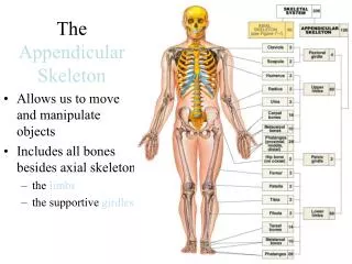
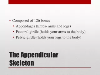
An Introduction to the Appendicular Skeleton • The Appendicular Skeleton • 126 bones • Allows us to move and manipulate objects • Includes all bones besides axial skeleton • The limbs • The supportive girdles
Figure 8-1 The Appendicular Skeleton SKELETAL SYSTEM 206 AXIAL SKELETON APPENDICULAR SKELETON 126 80 (see Figure 7–1) 2 Clavicle Pectoral girdle 4 Scapula 2 2 Humerus Upper limbs 60 2 Radius Ulna 2 Carpal bones 16 Metacarpal bones 10 28 Phalanges Pelvic girdle 2 Hip bone 2
Figure 8-1 The Appendicular Skeleton (Part 2 of 2) Femur 2 Lower limbs 60 Patella 2 Tibia 2 2 Fibula Tarsal bones 14 Metatarsal bones 10 28 Phalanges
8-1 The Pectoral Girdle • The Pectoral Girdle • Also called shouldergirdle • Connects the arms to the body • Positions the shoulders • Provides a base for arm movement • Consists of: • Two clavicles • Two scapulae • Connects with the axial skeleton only at the manubrium
8-1 The Pectoral Girdle • The Clavicles • Also called collarbones • Long, S-shaped bones • Originate at the manubrium (sternal end) • Articulate with the scapulae (acromial end)
Figure 8-2a The Right Clavicle Scapula Jugular notch Clavicle The position of the clavicle within the pectoral girdle, anterior view.
Figure 8-2b The Right Clavicle Acromial end Sternal end LATERAL MEDIAL Facet for articulation with acromion Superior view of the right clavicle.
Figure 8-2c The Right Clavicle Sternal facet Acromial end LATERAL MEDIAL Costal tuberosity Conoid tubercle Sternal end Inferior view of the right clavicle. Stabilizing ligaments attach to the conoid tubercle and the costal tuberosity.
8-1 The Pectoral Girdle • The Scapulae • Also called shoulder blades • Broad, flat triangles • Articulate with arm and collarbone • Anterior surface the subscapular fossa
8-1 The Pectoral Girdle • The Scapulae • Structures of the scapula • Body has three sides • Superior border • Medial border (vertebral border) • Lateral border (axillary border)
8-1 The Pectoral Girdle • The Scapulae • Body has three corners • Superior angle • Inferior angle • Lateral angle (head) • The scapular head • Holds glenoid cavity • Which articulates with humerus • To form shoulder joint (glenohumeral joint)
8-1 The Pectoral Girdle • The Scapulae • Processes of the glenoid cavity • Coracoid process • Anterior, smaller • Acromion • Posterior, larger • Articulates with clavicle • At the acromioclavicular joint
Figure 8-3a The Right Scapula Superior angle Acromion Superior border Coracoid process Lateral angle Subscapular fossa Body Lateral border Medial border Inferior angle Anterior view
Figure 8-3b The Right Scapula Supraglenoid tubercle Coracoid process Acromion Glenoid cavity Spine Lateral border Inferior angle Lateral view
Figure 8-3c The Right Scapula Supraspinous fossa Coracoid process Acromion Superior border Neck Spine Infraspinous fossa Body Medial border Lateral border Inferior angle Posterior view
8-1 The Pectoral Girdle • The Scapulae • Posterior features of the scapula • Scapular spine • Ridge across posterior surface of body • Separates two regions • Supraspinous fossa • Infraspinous fossa
8-2 The Upper Limbs • The Upper Limbs • Consist of: • The arms, forearms, wrists, and hands • Note: arm (brachium) = 1 bone, the humerus
8-2 The Upper Limbs • The Humerus • Also called the arm • The long, upper arm bone • Articulates with the pelvic girdle
8-2 The Upper Limbs • The Humerus • Tubercles of the proximal epiphysis • Separated by the intertubercular groove • Greater tubercle • Lateral • Forms tip of shoulder • Lesser tubercle • Anterior, medial
8-2 The Upper Limbs • The Humerus • Head • Rounded, articulating surface • Contained within joint capsule • Anatomical neck • Margin of joint capsule • Surgical neck • The narrow metaphysis
8-2 The Upper Limbs • The Humerus • The shaft • Deltoid tuberosity • A bulge in the shaft • Attaches deltoid muscle • Radial groove • For radial nerve • Posterior to deltoid tuberosity
8-2 The Upper Limbs • The Humerus • The distal epiphysis • Medial and lateral epicondyles • For muscle attachment • Condyle of the humerus • Articulates with ulna and radius
8-2 The Upper Limbs • The Humerus • Articular regions of the condyle • Trochlea • Coronoid fossa and olecranon fossa • Articulates with ulna • Capitulum • Radial fossa • Articulates with radius
Figure 8-4a The Right Humerus and Elbow Joint Head ad Greater tubercle Head Greater tubercle Lesser tubercle Anatomical neck Intertubercular groove Anatomical neck Surgical neck Surgical neck Deltoid tuberosity Deltoid tuberosity Radial groove Shaft Olecranon fossa Radial fossa Coronoid fossa Lateral epicondyle Lateral epicondyle Medial epicondyle Medial epicondyle Trochlea Trochlea Capitulum Anterior surface Condyle Posterior surface
Figure 8-4c The Right Humerus and Elbow Joint Humerus Medial epicondyle Trochlea Head of radius Capitulum Coronoid process of ulna Radial notch of ulna Elbow joint, anterior view
Figure 8-4d The Right Humerus and Elbow Joint Humerus Medial epicondyle Olecranon fossa Olecranon Trochlea of humerus Ulna Head of radius Elbow joint, posterior view
8-2 The Upper Limbs • The Forearm • Also called the antebrachium • Consists of two long bones • Ulna (medial) • Radius (lateral)
8-2 The Upper Limbs • The Ulna • The olecranon • Superior end of ulna • Point of elbow • Superior lip of trochlear notch • Articulates with trochlea of humerus • The coronoid process • Inferior lip of trochlear notch
8-2 The Upper Limbs • The Ulna • Articulations with the humerus • Forearm extended • Olecranon enters olecranon fossa • Forearm flexed • Coronoid process enters coronoid fossa
8-2 The Upper Limbs • The Ulna • Other articulations • Radial notch • Articulates with head of radius • Forms proximal radio-ulnar joint • Ulnar head • Prominent styloid process • Attaches to articular disc between forearm and wrist
Figure 8-5a The Right Radius and Ulna Olecranon Radial head Proximal radioulnar joint Neck of radius ULNA RADIUS Interosseous membrane Ulnar notch of radius Ulnar head Styloid process of ulna Styloid process of radius Posterior view
Figure 8-5b The Right Radius and Ulna Trochlear notch Coronoid process Radial head Radial notch Neck of radius Ulnar tuberosity Radial tuberosity ULNA RADIUS Interosseous membrane Distal radio-ulnar joint Ulnar head Styloid process of radius Anterior view
8-2 The Upper Limbs • The Ulna • Interosseous membrane • A fibrous sheet • Connects lateral margin of ulnar shaft to radius
8-2 The Upper Limbs • The Radius • Lateral bone of forearm • Disk-shaped radial head above the neck • Radial tuberosity below the neck, attaches biceps • Articulations of the radius • Ulnar notch • Distal end • Articulates with wrist and radius • Styloid process • Stabilizes wrist joint
Figure 8-5a The Right Radius and Ulna Olecranon Radial head Proximal radioulnar joint Neck of radius ULNA RADIUS Interosseous membrane Ulnar notch of radius Ulnar head Styloid process of ulna Styloid process of radius Posterior view
Figure 8-5b The Right Radius and Ulna Trochlear notch Coronoid process Radial head Radial notch Neck of radius Ulnar tuberosity Radial tuberosity ULNA RADIUS Interosseous membrane Distal radio-ulnar joint Ulnar head Styloid process of radius Anterior view
Figure 8-5c The Right Radius and Ulna Olecranon Trochlear notch Coronoid process Radial notch Ulnar tuberosity ULNA Lateral view of ulna, showing trochlear notch
8-2 The Upper Limbs • Eight Carpal Bones • Four proximal carpal bones • Four distal carpal bones • Allow wrist to bend and twist
8-2 The Upper Limbs • The Four Proximal Carpal Bones • Scaphoid • Near styloid process • Lunate • Medial to scaphoid • Triquetrum • Medial to lunate • Pisiform • Anterior to triquetrum
8-2 The Upper Limbs • The Four Distal Carpal Bones • Trapezium • Lateral • Trapezoid • Medial to trapezium • Capitate • Largest • Hamate • Medial, distal
Figure 8-6 Bones of the Right Wrist and Hand RADIUS RADIUS ULNA Lunate Lunate Scaphold Scaphold Triquetrum Trapezium Trapezium Pisiform Trapezoid Trapezoid I Capitate Capitate I Hamate V V IV Metacarpal bones IV II Metacarpal bones III III II Proximal phalanx Distal phalanx Proximal phalanx Middle phalanx Distal phalanx Posterior view Anterior view
8-2 The Upper Limbs • Metacarpal Bones • The five long bones of the hand • Numbered I–V from lateral (thumb) to medial • Articulate with proximal phalanges • Phalanges of the Hands • 14 total finger bones • Pollex (thumb) • Two phalanges (proximal, distal) • Fingers • Three phalanges (proximal, middle, distal)
Figure 8-6a Bones of the Right Wrist and Hand RADIUS ULNA Lunate Scaphold Triquetrum Trapezium Pisiform Trapezoid I Capitate Hamate V IV Metacarpal bones III II Proximal phalanx Distal phalanx Anterior view
Figure 8-6b Bones of the Right Wrist and Hand RADIUS ULNA Lunate Scaphold Triquetrum Trapezium Pisiform Trapezoid I Capitate Hamate V IV II Metacarpal bones III Proximal phalanx Middle phalanx Distal phalanx Posterior view
8-3 The Pelvic Girdle • The Pelvic Girdle • Made up of two hip bones (coxal bones) • Strong to bear body weight, stress of movement • Part of the pelvis • Coxal bones • Made up of three fused bones • Ilium (articulates with sacrum) • Ischium • Pubis
8-3 The Pelvic Girdle • Coxal Bones • The acetabulum • Also called the hip socket • Is the meeting point of the ilium, ischium, and pubis • Is on the lateral surface of the hip bone (coxal bone) • Articulates with head of the femur (lunatesurface) • Acetabular notch • A gap in the ridge of the margins of the acetabulum
Figure 8-7a The Right Hip Bone Ilium POSTERIOR ANTERIOR Ischium Pubis Iliac crest Anterior gluteal line Anterior superior iliac spine Posterior gluteal line Inferior gluteal line Posterior superior iliac spine Anterior inferior iliac spine Posterior inferior iliac spine Acetabulum Greater sciatic notch Acetabular notch Lunate surface of acetabulum Superior ramus of pubis Ischial spine Lesser sciatic notch Pubic tubercle Inferior ramus of pubis Ischial tuberosity Obturator foramen Ischial ramus Right hip bone, lateral view
8-3 The Pelvic Girdle • Marks of the Ilium • Greater sciatic notch • For sciatic nerve • Iliac crest • Upper brim • Iliac fossa • Depression between iliac crest and arcuate line