Download
1 / 31
320 likes | 541 Vues
Molecular Biology (MLMB-201). Department of Medical Laboratory Technology Faculty of Allied Medical Science. Lecturer: Dr. Mohamed Salah El-Din. Intended Learning Outcomes (ILO’s): Molecular biology course provides an overview of the molecular basis to cell structure and function.

E N D
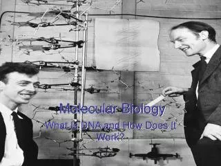
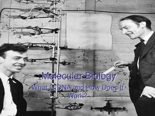
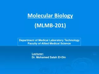
Molecular Biology (MLMB-201) Department of Medical Laboratory Technology Faculty of Allied Medical Science Lecturer: Dr. Mohamed Salah El-Din
Intended Learning Outcomes (ILO’s): • Molecular biologycourse provides an overview of the molecular basis to cell structure and function. • This course focuses on the structure, biosynthesis and function of DNA and RNA on the molecular level and how these interact among themselves and with proteins. Molecular biology techniques are essential for modern biological and medical research. This course will give you an introduction to DNA and RNA standard techniques. • Student will have basic knowledge of: • Cell organization. • DNA structure and function. • DNA Extraction. • RNA structure and function. • RNA Extraction. • Gene expression and protein biosynthesis. • Agarose gel electrophoresis for DNA/RNA; and SDS-PAGE for protein. • Polymerase Chain Reaction (PCR) – Theory, Types, Application. • Gene library and screening • DNA sequencing
Central Dogma Of Molecular Biology Chromosomes Thread-like structures present within the nuclei of cells. Chemically, a chromosome consists of an extremely long chain of DNA along with a coating protein. The combination of DNA and protein is called chromatin. The complete array of chromosomes in an organism is termed the karyotype.
A bit of DNA which acts as a code to control part of a cell's chemistry. Every human cell holds, within its nucleus, more than 50,000 different genes. Each gene is located at a particular position along the chromosome, termed locus. The particular form of the gene is termed the allele. What is a “gene”?
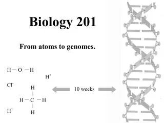
DNA STRUCTURE Structure of Nucleotide DNA RNA
Cytosine (C) Adenine (A) Guanine (G) Thymine (T) Thymine (T) Adenine (A) Guanine (G) Cytosine (C) Phosphoric Acid (Phosphate molecule) Deoxyribose (Sugar molecule)
Pyrimidine (T & C) Purine (A & G) nucleoside nucleotide
Watson-Crick model of a linear,double-stranded helix • A single polynucleotide strand of DNA is a copolymer in which deoxyribose and phosphate alternate; the side chains are single organic bases, one attached to each deoxyribose.
DNA FUNCTION DNA is the repository of genetic information in all organisms: (except for a relatively small number of viruses) • It must transmit its encoded information to the cell which contains it “Transcription” • It must replicate in order that a progeny cell has the same properties as its parent “Replication” • Evolutionary pressures have produced a mechanism for creating new information“Mutation” and for exchanging genes “Recombination”, so that new and better organisms can develop.
Mutation The copying process is not perfect, and very occasionally a fault occurs leading to a mutation (change) in the nucleic acid sequence; this, in turn, alters the structure of the DNA in one of the daughter cells-and thus leads to a change in one of its genes. Mutations may potentially occur at any site within the genome and may have several effects upon the structure and function of the genome. Recombination
DNA extraction Relevance ofDNAisolation Isolation of DNA is often the first step before further analysis: • DNA profiling • cloning • disease diagnosis • DNA sequencing • genetically modified organisms (GMO) -agriculture, pharmaceutical • Environmental testing, bioterrorism
Protocol Highlights:DNA Extraction • Collect cells from whole blood, tissues…etc. (NB. Ample cell collection is critical for success) • Add Lysis buffer to cells to break open cell and nuclear membranes and release nuclear contents • Digest sample with protease to degrade proteins • Precipitate DNA with cold alcohol in high salt
CH3 CH2 CH2 CH2 CH2 CH2 CH2 CH2 CH2 CH2 CH2 CH2 O O O S - O SDS Lysis Buffer:What is Lysis Buffer?• 50 mM Tris-HCI, pH 8.0• 1% SDSTris buffer to maintain the pH of the solution at a level where DNA is stable1% SDS to break open the cell and nuclear membranes, allowing the DNA to be released into the solution (SDS also denatures and unfolds proteins, making them more susceptible to protease cleavage).
Why Add Protease? • Protease is added to destroy nuclear proteins that bind DNA and cytoplasmic enzymes that breakdown and destroy DNA. • Protease treatment increases the amount of intact DNA that is extracted. Adding Salt • The protease solution already contains salt • Na+ ions of NaCI bind to the phosphate groups of DNA molecules, neutralizing the electric charge of the DNA molecules. • The addition of NaCI allows the DNA molecules to come together instead of repelling each other, thus making it easier for DNA to precipitate out of solution when alcohol is added. • Salts and buffers deactivate the enzymes that degrade DNA when released and stabilize the DNA (acid vs. base).
Adding Ice Cold Alcohol? • DNA does not dissolve in alcohol. • The addition of cold alcohol makes the DNA clump together and precipitate out of solution. • Precipitated DNA molecules appear as long pieces of fluffy, stringy, web-like strands. • Microscopic oxygen bubbles “aggregate” , or “fuse” together, as the DNA precipitates. • The larger, visible air bubbles “lift” the DNA out of solution, from the aqueous into the organic phase.
DNA extraction DNA is easy to prepare and store because DNAases are easy destroyed by heating. • Phenol/chloroform/isoamyl alcohol extraction: • Phenol is organic solvent. • Nucleic acids prefer aqueous solutions. • But, proteins, lipids and carbohydrates prefer organic environment. • So, after addition of phenol to the cell lysate, the reaction tube will have two phases: • Upper aqueous layer containing DNA. • Lower organic layer containing phenol + proteins.
Salting out method: Cell lysis. Protein digestion by proteinase enzyme. Protein precipitation by high salt concentration. Salt also reduces the repulsion of the negatively charged DNA molecules. Centrifugation will remove the precipitated proteins. The supernatant contains the DNA. DNA is then precipitated by adding ethanol. The precipitated DNA is resuspended in the desired buffer.
Spin column: Commercial DNA extraction kits are based on selective adsorption of DNA to silica gel membrane columns or glass fiber matrix columns. Binding of DNA to silica or glass fiber is enhanced by high salt concentration. The bound DNA will then be eluted by elution buffer with low salt concentration.
Plasmid extraction • Alkaline lysis method: • Cell lysis. • Then, we add sodium hydroxide. • The alkaline pH will denaturate chromosomal DNA but not the plasmid DNA. • Then we add sodium or potassium acetate that will neutralize pH. • So, chromosomal DNA will renature and aggregate with proteins. • By centrifugation, proteins + chromosomal DNA will precipitate while plasmid DNA will remain in the supernatant.
Quantification • Quantification of extracted nucleic acids is done by using spectrophotometer as follows: • At wave length 260: optical density (OD) of 1 means that: • The concentration of DNA= 50 µg/ml • The concentration of RNA= 40 µg/ml • So, the concentration of extracted DNA in a sample= OD at 260 x 50 x dilution factor
DETERMINING DNA CONCENTRATION AND PURITY BY SPECTROPHOTOMETRY • PROCEDURE: • Fill two cuvettes with TE buffer. Read and record the A260 of the sample cuvette against the blank. Repeat at 280 nm. • 2. Dilute the DNA in 400 µl of TE such that the A260 is ideally between 0.1 to 1.0. Mix well. • 3. Empty and clean the sample cuvette and add the diluted DNA. • 4. Record absorbance of DNA sample at both 260 and 280 nm. Correct the readings as necessary using the blank values you determined in step 1. • 5. The absorbance at 260 nm allows calculation of the concentration of DNA or RNA in the sample. An OD of 1 corresponds to approximately 50 ng/µl for double-stranded DNA, 40 ng/µl for RNA, and 32 to 34 ng/µl for single-stranded DNA and typical oligonucleotides. The A260/A280 ratio can provide a very rough estimate of the purity of the nucleic acid. Relatively pure preparations of DNA and RNA have A260/A280 values of 1.8 and 2.0, respectively. Phenol contamination will result in significantly lower A260/A280 ratios. Such contamination makes accurate quantitation of DNA or RNA impossible. Note however that the A260/A280 ratio can not be used to determine whether there is significant protein contamination in a nucleic acid preparation (Glasel, J. Biotechniques 18:62 (1995).
It is very important to assess purity of the extracted DNA (the degree of protein contamination). Purity is assessed by: Determine the ratio between the OD of the sample at 260 nm and 280 nm. (OD 260/OD 280). If the ratio is 2: this means that protein contamination is zero. If the ratio is < 2:this means protein contamination of the extracted DNA. N.B. Notice that the concentration of the extracted nucleic acid in a given sample can be roughly estimated by observing the fluorescence intensity of the band obtained on agarose gel after electrophoresis.
Now You Can Capture Your Unique Essence! & Prepare DNA Necklaces The DNAin the glass vial can last for years. Add more alcohol into the vial if some evaporation occurs. Congratulations! You have just created your very own DNA Necklace!
Assignment: As a part of the semester activity, one student is selected every week to prepare a short seminar about his/her point of interest in one of the lecture topics. That to be discussed and evaluated during the next lecture.