Download
1 / 20
200 likes | 224 Vues
This guided notes resource provides detailed information on the three parts of the axial skeleton: the skull, vertebral column, and bony thorax. Learn about the composition, functions, and unique characteristics of each component, from the cranial bones to the different types of ribs. Explore how the axial skeleton supports the body and protects vital organs. Enhance your understanding with clear explanations and valuable insights. Perfect for students studying anatomy or anyone interested in the human skeletal system.

E N D
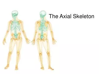
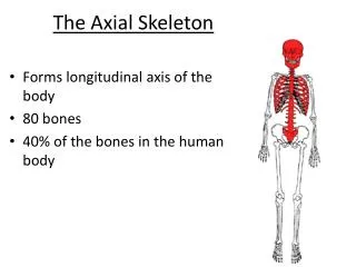
1. The axial skeleton can be divided into three parts, which are the skull, the vertebral column, and the bony thorax.
2. The skull is formed by two sets of bones. The cranium encloses and protects the fragile brain tissue. The facial bones hold the eyes in an anterior position.
14 bones compose the face. Twelve are paired; only the mandible and vomer are single.
5. The two maxilla fuse to form the upper jaw. Like many other facial bones, the maxillae contain sinuses, which drain into the nasal passages. They are called paranasal sinuses.
6. The zygomatic bones are commonly referred to as the cheekbones.
7. The mandible, or lower jaw, is the largest and strongest bone of the face. It joins the temporal bones on each side of the face, forming the only freely movable joints in the skull.
8. The hyoid bone is unique in that it is the only bone of the body that does not articulate directly with any other bone. The hyoid bone serves as a movable base for the tongue and attachment point for neck muscles.
9. In the newborn, the skull has regions that have yet to be converted to bone. These fibrous membranes are called fontanels; they connect the cranial bones. The fontanels allow the fetal skull to be compressed slightly during birth. They can no longer be felt 22 to 24 months after birth.
10. The vertebral column extends from the skull to the pelvis, where it transmits the weight of the body to the lower limbs. The spine is formed from 26 irregular bones connected and reinforced by ligaments in such a way that a curved, flexible structure results.
11. The single vertebrae are separated by pads of flexible fibrocartilage called intervertebral discs, which cushion the vertebrae and absorb shocks.
Common Structures of Vertebrae • Body: disc-like; weight-bearing part of vertebra • Vertebral Arch: formed from the joining of the laminae and pedicles • Vertebral Foramen: canal through which the spinal cord passes • Transverse Process: (2) lateral projection from the vertebral arch • Spinous Process: single projection arising posteriorly from vertebral arch • Articular Processes: paired projections lateral to the foramen, allowing vertebrae to form joints
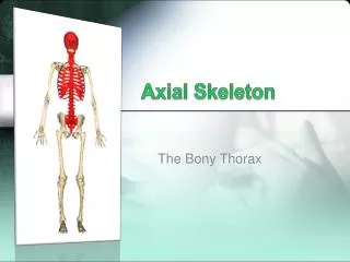
14. The sternum, ribs, and thoracic vertebrae make up the bony thorax.
15. The sternum is a typical flat bone, and is a result of the fusion of the manubrium, body, and xiphoid process.
16. Twelve pairs of ribs form the walls of the thoracic cage. All the ribs articulate with the vertebral column and then curve downward and toward the anterior body surface.
3 Different Types of Ribs • True ribs: the first 7 pairs, which attach directly to the sternum by costal cartilage • False ribs: the next 5 pairs attach indirectly to the sternum by a shared cartilage • Floating ribs: do not attach to the sternum at all. They are the lowest 2 pairs of false ribs.