Download
1 / 32
340 likes | 1.36k Vues
Learn about cirrhosis of the liver, its possible causes, clinical signs, and drug treatments, including alcohol withdrawal management. Explore Mrs. MW's case and recommended interventions for her condition.

E N D
Pharmaco-therapeutics 2 Prince Sattam Bin AbdulAziz University College Of Pharmacy Cirrhosis • Mohammad Ruhal Ain • R Ph, PGDPRA, M Pharm (Clin. Pharm) • Department of Clinical Pharmacy • E-mal: m.alain@sau.edu.sa Email : m.alain@sau.edu.sa
Liver cirrhosis case • Mrs MW, 59 years old, is divorced and unemployed. She was admitted to an acute medical ward at the hospital presenting with general malaise, a grosslydistended abdomen, swollen ankles and jaundice. It was also noted that she smelt of alcohol and was showing signs of alcohol withdrawal
1 What is cirrhosis of the liver? • 2 List possible causes of cirrhosis. • 3 What other clinical signs and symptoms may Mrs MW present with? • 4 What drug treatment, including dose, would you recommend for Mrs MW’s • alcohol withdrawal? What recommendations would you make if the patient was • unable to take the medication orally?
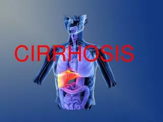
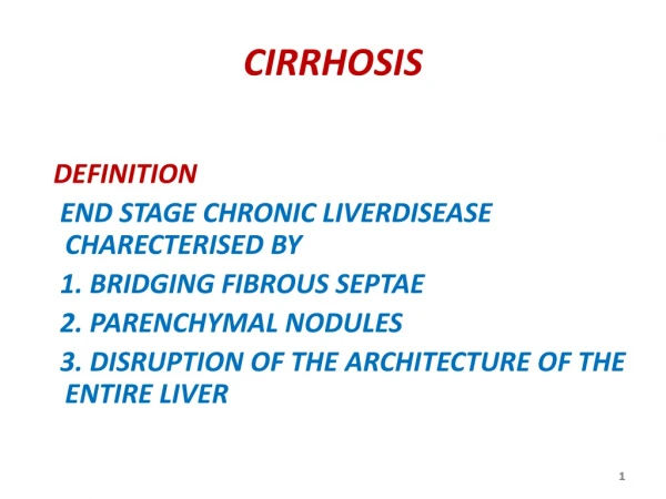
What is cirrhosis of the liver? • Cirrhosis is defined as the histological development of regenerative nodules surrounded by fibrous bands in response to chronic liver injury. • It is an advanced stage of liver fibrosis that is accompanied by distortion of the hepatic vasculature.
List possible causes of cirrhosis • Causes of cirrhosis can usually be identified by the patient’s history combined with serological and histological investigation. Alcoholic liver disease and hepatitis C and B are the most common causes of cirrhosis • The association of excessive alcohol consumption with liver disease has been recognised for centuries. • After the identification of the hepatitis C virus and of non-alcoholic steatohepatitis in obese patients with diabetes, the diagnosis of cirrhosis without an apparent cause (cryptogenic cirrhosis) is rarely made. • Genetic causes of cirrhosis include haemochromatosis and Wilson’s disease. • Epidemiological studies have identified a number of factors that contribute to the risk of developing cirrhosis. • Regular (moderate) alcohol consumption, age older than 50 years, and male gender are examples that increase cirrhosis risk in chronic hepatitis C infection, and older age, obesity, insulin resistance or type 2 diabetes, hypertension and hyperlipidaemia in non-alcoholic steatohepatitis.
What other clinical signs and symptoms may Mrs MW present with? • Cirrhosis is often asymptomatic until complications of liver disease are present. • Mrs MW may present with itching, jaundice, dark urine, pale fatty stools, • abdominal pain, nausea, fatigue, bleeding – such as nose bleeds, hepatic encephalopathy, hepatomegaly, ascites, distended abdominal veins, spider angiomata, palmar erythema and asterixis. She may also present with the signs and symptoms of alcohol withdrawal, which include irritability, anxiety, tachycardia, tremor, sweating, confusion and hallucinations.
What drug treatment, including dose, would you recommend for Mrs MW’s alcohol withdrawal? What recommendations would you make if the patient was unable to take the medication orally? • Long-acting benzodiazepines (e.g. diazepam and chlordiazepoxide) are used to attenuate alcohol withdrawal symptoms but they also have a dependence potential. • To minimise the risk of dependence, administration should be for a limited period only (e.g. chlordiazepoxide 20 mg 4 times daily, gradually reducing to zero over 7–14 days). • Mild alcohol withdrawal symptoms may be treated with a lower starting dose, such as 15 mg four times a day. • In all cases, the patient should be counselled about the proposed length of the treatment course. • Benzodiazepines should not be prescribed if the patient is likely to continue drinking alcohol. In patients unable to take medication by the oral route, diazepam may be administered by intramuscular or slow intravenous injection (into a large vein, at a rate of not more than 5 mg/min), at a dose of 10 mg, repeated if necesary after not less than 4 hours. Alternatively, diazepam may be administered via the rectal route as a rectal solution or suppository. The intramuscular route should only be used when both the oral and intravenous routes are not possible.
Mrs MW weighs 61 kg (with the ascites) and her laboratory data are as follows: • Total protein 49 g/L (63–80 g/L) • Albumin 20 g/L (32–50 g/L) • Total bilirubin 114 micromol/L (<17 micromol/L) • ALP 382 IU/L (100–300 IU/L) • ALT 88 IU/L (5–42 IU/L) • INR 1.6 • GGT 306 IU/L (<50 IU/L) • Diagnosis of alcoholic cirrhosis of the liver was made based on Mrs MW’s clinical features, liver function tests, abdominal ultrasound, CT scan and liver biopsy.
1. Describe how Mrs MW’s laboratory results relate to her diagnosis. • 2. What treatment would you recommend to reduce her risk of bleeding? • 3. What medications would you advise Mrs MW to avoid in view of her bleeding risk? • 4 What treatment options are available to help Mrs MW abstain from alcohol in the future?
Describe how Mrs MW’s laboratory results relate to her diagnosis. • Albumin is synthesised in the liver and has a long half-life of around 20 days. • Mrs MW’s low serum albumin is indicative of chronic liver disease. • Albumin synthesis is also depressed in states of poor nutrition. • Bilirubin is carried within the plasma by albumin to the liver, where it is conjugated by glucuronidation. • Subsequently, low circulating albumin levels and/or damage to the liver cells result in reduced conjugation of the bilirubin.
Blockage of the biliary tract (e.g. cholestasis) will result in increased levels of conjugated bilirubin. • Bilirubin levels of more than 35 micromol/L can result in visual signs of jaundice. • The concentration of both the unconjugated and conjugated bilirubin are likely to be raised in Mrs MW. However, only total bilirubin was measured in this case. • Alkaline phosphatase (ALP) is present in high concentrations in the cells lining the biliary tract and an ALP level exceeding 300 IU/L, together with a raised bilirubin as in the case of Mrs MW, is indicative of cholestasis. Jaundicebecomes progressively more severe in unrelieved cholestasis. • Alanine aminotransferase (ALT) is present in high concentrations in the liver and the enzyme is released during hepatocellular damage and a modestly raised level like Mrs MW’s is indicative of chronic liver disease.
The increased susceptibility to bleeding observed in patients with liver failure (raised INR) results from depressed fibrinogen levels and the reduced synthesis of clotting factors by the cirrhotic liver. • In addition, the absorption of fat-soluble vitamin K is impaired in cholestasis and subsequently the synthesis of vitamin K-dependent clotting factors is reduced. • Gamma-glutamyl transferase (GGT) is found in the liver, but this test is relatively non-specific. It is released following tissue damage and is raised in cholestasis in parallel with ALP. GGT release is stimulated by alcohol and some drugs (such as phenytoin and carbamazepine), and therefore the GGT level can be used to assess abstinence in alcoholics, like Mrs MW.
What treatment would you recommend to reduce her risk of bleeding? • Mrs MW has a raised INR value of 1.6 and is therefore at increased risk of bleeding. • Vitamin K can be given to correct the vitamin deficiency, either as an intravenous injection or orally. Konakion MM is phytomenadione (10 mg/mL) in amixed micelles vehicle. • Konakion MM may be administered by slow intravenous injection or by infusion in glucose 5%. • For oral administration, the water-soluble preparation menadiol sodium phosphate, is used in patients with hepatic disease, especially biliary obstruction. • The usual dose is 10 mg daily. Alternatively, phytomenadione tablets may be used in those patients who do not have impaired fat absorption. • If vitamin K is ineffective in controlling the clotting times (monitored via the INR result) then fresh frozen plasma may be required.
What medications would you advise Mrs MW to avoid in view of her bleeding risk? • Medications known to increase the risk of bleeding in cirrhotic patients include • aspirin, clopidogrel, dipyridamole, corticosteroids, NSAIDs, heparin and warfarin. • Mrs MW would need to be counselled about the risks associated with these medications and advised to always check with the pharmacist before buyingany medications over the counter
What treatment options are available to help Mrs MW abstain from alcohol in the future? • Disulfiram is an aversive therapy that works by inhibiting acetaldehyde dehydrogenase. • Interactions between disulfiram and alcohol can result in potentially severe reactions, such as myocardial infarction, congestive heart failure, respiratorydepression and death. • Patients taking disulfiram should be warned of the possible presence of alcohol in liquid medicines, tonics, foods and even in toiletries and mouthwashes. • Patient adherence to disulfiram is poor and there is a lack of strong evidence for its effectiveness, thus it is not routinely recommended. • Acamprosate is indicated for the maintenance of abstinence in alcohol dependent adults. • It appears to decrease brain hyper excitability during alcohol withdrawal, which may reduce alcohol consumption. • Treatment should be initiated as soon as possible after the alcohol-withdrawal period is complete. • The recommended period of treatment with acamprosate is one year and treatment should be combined with counselling. • The GGT level can be monitored as a marker of abstinence from alcohol.
Mrs MW, 59 years old, is divorced and unemployed. She was admitted to an acute medical ward at the hospital presenting with general malaise, a grossly • distended abdomen, swollen ankles and jaundice. It was also noted that she smelt of alcohol and was showing signs of alcohol withdrawal. • On examination, Mrs MW was found to be encephalopathic. The doctors decided to treat her encephalopathy and ascites.
What is hepatic encephalopathy? What are the clinical signs and symptoms? • 2 What factors may precipitate hepatic encephalopathy? • 3 List two treatment options for the management of Mrs MW’s hepatic encephalopathy. Describe the mechanism of action for one of these. • 4 What factors are likely to have contributed to the development of ascites in Mrs MW? • 5 Name two treatment options for the management of Mrs MW’s ascites. Describe the pharmacology of these therapies, including any potential side-effects. What would you monitor in order to determine whether the therapy was effective?
What is hepatic encephalopathy? What are the clinical signs and symptoms? • Hepatic encephalopathy is a neuropsychiatric syndrome which may complicate almost all types of liver disease. • It may occur intermittently and be reversible or may occur acutely, with rapid progression to coma and death. • Mrs MW presented with signs of hepatic encephalopathy including flapping tremor of the hands, intellectual deterioration, slurred speech, confusion, drowsiness and irritability.
2 What factors may precipitate hepatic encephalopathy? • Factors that may precipitate hepatic encephalopathy include: • hypokalaemia and/or profound diuresis caused by a brisk response to a potent diuretic, • diarrhoea and vomiting, because of the resulting fluid and electrolyte imbalance, constipation, • a large protein meal or gastrointestinal haemorrhage, infection, especially peritonitis, • and CNS depressant drugs, such as opioids or benzodiazepines.
List two treatment options for the management of Mrs MW’s hepatic encephalopathy. Describe the mechanism of action for one of these. • Treatment goals for hepatic encephalopathy include provision of supportive care, identification and removal of precipitating factors, reduction in the nitrogenous load from the gut and optimisation of long-term therapy.
Therapy should be directed toward improving mental status via bowel cleansing with lactulose or with enemas. The dose of lactulose should be titrated to give two soft stools per day without diarrhoea. • Lactulose is metabolised in the colon to lactic, acetic and formic acids, causing the pH of the colon to drop from 7 to 5. • Colonic acidification with lactulose alters the bacterial population and favours the growth of weak ammonia-producing bacteria rather than proteolytic ammonia producers such as E. coli. • In addition, the drop in colon pH leads to ionisation of nitrogenous products with a subsequent reduction in their absorption from the gastrointestinal tract into the blood.
Third, the osmotic laxative effect speeds the intestinal transit, thereby decreasing the time available for the absorption of potentially toxic nitrogen compounds. It is imperative to monitor • Mrs MW’s bowel frequency while treating with lactulose since fluid and electrolyte • imbalances secondary to diarrhoea may precipitate hepatic encephalopathy. • Mrs MW’s mental test score should also be repeated to assess benefit. • Neomycin (4 g daily in divided doses) is a non-absorbable antibiotic which may be used to reduce the number of bacteria in the bowel that normally break down protein. • Neomycin therapy is usually limited to one week’s therapy because some absorption may occur with a risk of nephrotoxicity and ototoxicity.
What factors are likely to have contributed to the development of ascites in Mrs MW? • Mrs MW presented with a swollen abdomen, swollen ankles, pitting oedema and breathlessness. There are two key factors involved in the pathogenesis of ascites formation, namely, sodium and wate retention and portal hypertension.
The development of renal vasoconstriction in cirrhosis is partly a homeostatic response involving increased renal sympathetic activity and activation of the renin–angiotensin system to maintain blood pressure during systemic vasodilatation. • Decreased renal blood flow decreases glomerular filtration rate and thus the delivery and fractional excretion of sodium.
Cirrhosis is associated with enhanced reabsorption of sodium both at the proximal tubule and at the distal tubule. Increased reabsorption of sodium in the distal tubule is due to the increased circulating concentrations of aldosterone, occurring secondary to the reduced hepatic metabolism of aldosterone. • However, some patients with ascites have normal plasma concentrations of aldosterone, leading to the suggestion that sodium reabsorption in the distal tubule may be related to enhanced renal sensitivity to aldosterone.
In compensated cirrhosis, sodium retention can occur in the absence of vasodilatation and effective hypovolaemia. Sinusoidal portal hypertension can reduce renal blood flow even in the absence of haemodynamic changes in the systemic circulation, suggesting the existence of a hepatorenal reflex. • Portal hypertension increases the hydrostatic pressure within the hepatic sinusoids and favours transudation of fluid into the peritoneal cavity. Systemic vasodilatation, the severity of liver disease and portal pressure contribute to the abnormalities of sodium handling in cirrhosis.
Name two treatment options for the management of Mrs MW’s ascites. Describe the pharmacology of these therapies, including any potential side-effects. What would you monitor in order to determine whether the therapy was effective? • Management of ascites aims to mobilise ascitic fluid, relieve abdominal discomfort • and breathlessness and to exclude infection. Diuretics have been the mainstay • of treatment of ascites since the 1940s when they first became available.
Spironolactone, an aldosterone antagonist, is the drug of choice since • secondary hyperaldosteronism often coexists in patients with hepatic ascites. • Aldosterone is usually metabolised by the liver and is highly protein bound, therefore the free aldosterone levels are raised in cirrhosis. Spironolactone competes with aldosterone for receptor sites in the distal tubule, resulting in potassium retention and sodium and water loss. • The initial dose of spironolactone is 100–200 mg and can be slowly increased according to response. There is a lag of 3–5 days between the beginning of spironolactone treatment and the onset of the natriuretic effect.
The aim is to remove the fluid gradually with a maximum weight loss of • 0.5 kg/day in the absence of peripheral oedema, or 1.0 kg/day if peripheral • oedema is present. Too rapid a diuresis will result in intravascular fluid loss rather than the peripheral oedema. • The diuretic should be stopped if the serum sodium falls below 120 mmol/L or if there is a rising serum creatinine. Urinary electrolytes should be monitored to ensure that the spironolactone therapy is effective. • The aim is to reverse the sodium/potassium ratio in the urine so that • more sodium than potassium is excreted. Most frequent side-effects of spironolactone are those related to its anti-androgenic activity, such as decreased libido, impotence and gynaecomastia in men and menstrual irregularities in women. • Other side-effects include hyperkalaemia, uraemia, hyponatraemia and nausea.
In addition to spironolactone, ascites can be managed by paracentesis. • That is the removal (‘tapping’) of ascitic fluid from the peritoneal cavity under aseptic conditions. • A colloid (human albumin solution (20%)) is infused (40 Ml (8 g of albumin) per litre of ascites drained) intravenously during paracentesis, in order to prevent intravascular volume depletion and the onset of renal failure. • Following paracentesis, ascites recurs in the majority (93%) if diuretic therapy is not reinstituted, but recurs in only 18% of patients treated with spironolactone. • Cirrhotic ascites can become infected and if peritonitis occurs • survival depends on early, vigorous antibiotic therapy. The patient should be monitored for signs of infection