Download

1 / 50
500 likes | 569 Vues
Explore the morphological characteristics, classification, etiological factors, and pathogenesis of cirrhosis, an end-stage liver disease, with detailed insights into clinical features. Understand the complexities to tackle liver health effectively.

E N D
CIRRHOSIS Dr Ramadas nayak Professor & hod pathology
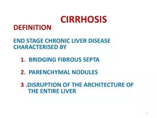
CIRRHOSIS • Definition: • Cirrhosis is an end stage of any chronic liver disease. • It is a diffuse process (entire liver is involved) • characterized by fibrosis and • conversion of normal architecture to structurally abnormal regenerating nodules of liver cells.
CIRRHOSIS • Morphological Characteristics • The three main morphologic characteristics of cirrhosis are: • 1. Fibrosis • 2. Regenerating Nodules • 3. Loss of Architecture
CIRRHOSIS • Morphological Characteristics • 1. Fibrosis • It is the characteristic feature of progressive liver damage. • The fibrous tissue form delicate bands or broad scars and link portal tracts with one another and portal tracts with terminal hepatic veins.
CIRRHOSIS • Morphological Characteristics • 2. Regenerating Nodules • Liver cell damage is compensated by regeneration of hepatocytes. • These regenerating hepatocytes forms nodules and are surrounded by fibrosis. • Nodularity results from cycles of hepatocyte regeneration and scarring. • The regenerating liver cells does not maintain the normal architecture. • The size of nodules vary from very small (< 0.3 cm, micronodules) to large (several centimeters, macronodules).
CIRRHOSIS • Morphological Characteristics • 3. Loss of Architecture • The hepatocyte injury and consequent fibrosis are diffuse processes, which occur in the entire liver. • This disrupts the architecture of the entire liver.
CIRRHOSIS • Classification • Morphological Classification (Fig. 14.20) • Depending on the size of the regeneration nodules cirrhosis is classified • Micronodular Cirrhosis • Macronodular Cirrhosis • Mixed Cirrhosis
CIRRHOSIS • Classification • Morphological Classification • Micronodular Cirrhosis • It is characterized by regular and small nodules measuring less than 3 mm in diameter. • The fibrous tissue septa are usually thin and fibrous septa bridge portal tracts and central veins (portal-portal and/portal-central) resulting in a small nodule without central structures (e.g. alcoholic cirrhosis).
CIRRHOSIS • Classification • Morphological Classification • Macronodular Cirrhosis • It is characterized by the presence of nodules of variable size, more irregular than in the micronodular cirrhosis and usually larger than 3 mm in diameter. • The fibrous tissue septa are broad and the nodules are more variable in composition and are often composed of multiple acini. • Micronodular cirrhosis can be converted into a macronodular form continued regeneration and expansion of existing nodules. • For example cirrhosis associated with chronic hepatitis. The macronodular cirrhosis has an increased risk of developing carcinoma of liver.
CIRRHOSIS • Classification • Morphological Classification (Fig. 14.20) • Mixed Cirrhosis • It consists of both micronodules and some macronodules. • Depending on the activity, each of these forms may sub-classified as an active and inactive form.
CIRRHOSIS • Etiological Classification • This classification takes into consideration clinical, biochemical, immunological or biopsy features • Main causes of cirrhosis • Alcohol is one of the commonest causes • Viral hepatitis (HBV and HCV) • Non-alcoholic steatohepatitis (NASH) • Hemochromatosis • Autoimmune liver disease (autoimmune hepatitis and primary biliary cirrhosis) • Intrahepatic or extrahepatic biliary obstruction: Recurrent biliary obstruction (e.g. gallstones) • Metabolic disorders: Wilson’s disease • ‘Cryptogenic’ (hidden cause) or idiopathic • Drugs and toxins • Indian childhood cirrhosis.
CIRRHOSIS • Pathogenesis • Four important processes are involved: • Death of liver cells with loss of architecture: • Fibrosis • Regenerating nodules • Vascular reorganization
CIRRHOSIS • Pathogenesis • Four important processes are involved: • Death of liver cells with loss of architecture: • The pathogenesis of hepatocyte injury varies depending on the etiological agent.
CIRRHOSIS • Pathogenesis • Fibrosis • It is mainly due to the activation of hepatic stellate cells, which are transformed into highly fibrogenic cells called myofibroblasts. • Regenerating nodules • The liver cell damage and fibrosis stimulate the surviving hepatocytes to regenerate and proliferate to form regenerating nodules • •Vascular reorganization: The parenchymal damage and fibrosis disrupt the vascular architecture of the liver
Morphology • Loss of architecture • Fibrosis • Regenerating nodules
CIRRHOSIS • Clinical Features • The clinical features of cirrhosis range widely: • Initial phase: It is termed as “compensated” cirrhosis, the patient may be asymptomatic. • Later phase: It is termed as “decompensated” cirrhosis, presents with complications of portal hypertension or liver dysfunction (or both).
CIRRHOSIS • Clinical Features • Nonspecific clinical manifestations: Anorexia, weight loss, weakness, and in advanced disease, symptoms and signs of hepatic failure may develop. • Hepatic failure is usually precipitated by systemic infection or gastrointestinal hemorrhage.
CIRRHOSISPortal Hypertension • Portal hypertension is defined as the elevation of the hepatic venous pressure above 7 mm Hg. • Causes of Portal Hypertension • The causes of portal hypertension can be divided into three categories: • Prehepatic causes: Obstructive thrombosis of the portal vein before it ramifies within the liver or massive splenomegaly with increased splenic vein blood flow. • Posthepatic causes: E.g. severe right-sided heart failure, constrictive pericarditis, and hepatic vein outflow obstruction. • Intrahepatic causes: E.g. cirrhosis (main cause), schistosomiasis, massive fatty change and diffuse fibrosing granulomatous disease (e.g. sarcoidosis).
CIRRHOSIS • Pathogenesis of Portal Hypertension in Cirrhosis • It is produced by a combination of two simultaneously occurring processes: • 1. Increased intrahepatic resistance to blood flow through the liver: • 2. Increase in portal venous inflow (flow):
CIRRHOSIS • Pathogenesis of Portal Hypertension in Cirrhosis • It is produced by a combination of two simultaneously occurring processes: • 1. Increased intrahepatic resistance to blood flow through the liver: • Deposition of fibrous tissue (scarring) • Fibrosis and compression by regenerative nodules (fixed component) increases the sinusoidal vascular resistance. • In the fibrous septa, anastomosis develop between the arterial and portal system. • These impose arterial pressures on the low pressure portal venous system and contribute to portal hypertension.
CIRRHOSIS • Pathogenesis of Portal Hypertension in Cirrhosis • It is produced by a combination of two simultaneously occurring processes: • 2. Increase in portal venous inflow (flow): • It results from the hyperdynamic circulation/consequences of portal hypertension
CIRRHOSIS • Consequences of Portal Hypertension • i. Ascites. • ii. Formation of portosystemic venous shunts. • iii. Congestive splenomegaly. • iv. Hepatic encephalopathy.
CIRRHOSIS • Ascites • Ascites is defined as the accumulation of excess fluid in the peritoneal cavity. • The most common (85% of cases) cause of ascites is portal hypertension caused by cirrhosis. • Pathogenesis of Ascites in Cirrhosis • It is complex process and involves the following mechanisms: • Portal hypertension: It increases the hydrostatic pressure in portal vein. • Hypoalbuminemia: It is due to decreased synthetic function in a cirrhotic liver → reduces the plasma oncotic pressure. • Splanchnic vasodilation and hyperdynamic circulation. Percolation of hepatic lymph into the peritoneal cavity: In cirrhosis, hepatic lymphatic flow exceeds thoracic duct capacity. The excess lymph may percolate (pass through liver) into the peritoneal cavity and cause ascites.
CIRRHOSIS • Portosystemic Shunts/Varices and Variceal Hemorrhage • Main Sites of Portosystemic Shunting/Bypasses • Main sites are • •Esophagogastric junction: These collaterals produce gastroesophageal varices. • •Veins around and within the rectum: It results in rectal varices (manifest as hemorrhoids). • •Retroperitoneum: Collaterals may form in the retroperitoneum, especially in females and communicate between the ovarian vessels and iliac veins. • •Umbilicus: Produce prominent collaterals around the umbilicus. They appear as dilated subcutaneous veins extending from the umbilicus toward the rib margins (caput medusae).
CIRRHOSIS • Splenomegaly and Hypersplenism • Congestive splenomegaly. The spleen is enlarged and varies in size. • Massive splenomegaly may give rise to the syndrome of hypersplenism. • Hypersplenism is characterized by a decrease in the lifespan of all of the formed elements of the blood (pancytopenia). • Pancytopenia of hypersplenism is due to the prolonged transit time of blood through the hyperplastic spleen.
CIRRHOSIS • Morphology • Gross: The spleen is firm and enlarged; up to 1000 g. • Cut surface is uniformly deep red. • Microscopy: The spleen shows dilated sinusoids with thickening of wall due to fibrous tissue. • Focal areas of hemorrhages may lead to the formation of fibrotic, iron-laden nodules. • These are known as Gamna-Gandy bodies.
CIRRHOSIS • Endocrine Complications are Associated with Cirrhosis • It may be either due to the direct effects of alcohol abuse or hepatic dysfunction. • In Men • Hyperestrogenism • Chronic liver failure → reduced hepatic catabolism of estrogens + weak androgens are converted to estrogenic compounds in peripheral tissues → hyperestrogenism → leads to feminization. • The portosystemic shunts secondary to portal hypertension in cirrhosis allow these hormones to bypass the liver. • Feminization is characterized by gynecomastia, a female body habitus, and a female distribution of pubic hair. Hyperestrogenism also causes vascular manifestations, which include spider angiomas (upper trunk and face) and palmar erythema. • Hypogonadism • Chronic alcoholics also develop hypogonadism, which is manifested as testicular atrophy, impotence, and loss of libido. These are due to direct toxic action of alcohol.
CIRRHOSIS • Endocrine Complications are Associated with Cirrhosis • In Women • They may show features of gonadal failure, presenting as oligomenorrhea, amenorrhea, infertility, ovarian atrophy, and loss of secondary sex characteristics. • These effects are due to direct toxic action of alcohol on gonads.