Head Injury
770 likes | 946 Vues
This document provides an extensive examination of head injuries, including historical trepanation techniques, classifications of brain injuries by Petit (1774), and detailed analysis of various types of head injuries such as cerebral concussion, contusion, and compression. It discusses the causes of head injuries in different populations, including traffic accidents and falls. The text reviews clinical presentations, severity ratings using the Glasgow Coma Scale, and the pathogenesis, pathology, and treatment options available for different forms of head trauma.

Head Injury
E N D
Presentation Transcript
Prehistorycal types of trepanation • 1-вишкрібання • 2-проскрібування канавки • 3-пробуравлення і вирізання • 4-шляхом прямокутних розрізів
Classification of Brain Injury, Petit, 1774 • Cerebral concussion (commotio cerebri) • Cerebral contusion(contusio cerebri) • Cerebral compression(compresio cerebri)
Causes of death in different age groups other cardiovascular diseases trauma neoplasms head injury
Occurrence of head injury in male and female Male Female
Causes of head injury in the USA • Fall from e height • Trafic accidents
Males Compression Contusion Concussion Females Compression Contusion Concussion Structure of types of head injury in different age groups and sex
On pathology basis • focal • diffuse
depending on infection risk • Closed • Open • penetrating • not penetrating
Clinical forms of head injury • Cerebral concussion • Brain contusion • Mild • moderate • severe • Diffuse axonal injury • Cerebral compression • Head compression
Initial lesions contusion diffuse axon injury hemorrhages injury of cranial nerves Secondary lesions Intracranial cerebral compression with hematomas Vioaltion of CSF and blood circulation Brain edema Extracranial Anemia hypoxemia hypertermia Pathogenesis of head injury
Clinical presentations of head injury • Signs of injury on the scalp (wounds, contusion) • Impaired consciousness • Amnesia • Focal neurological deficit • Pupil asymmetry • Cranial nerve deficit • Paresis • Reflex asymmetry and depression • Aphasia • Seizures
Level of consciousness • Clear consciousness - full and adequate orientation and reactions. Possible amnesia. • Mild– slight sleepiness,some time and place disorientantion, some slowness in command obey, • – hypersomnia,disorientation, only elementary verbal contact is possible, obeys only simplest verbal instructions. • Stupor – verbal contact is impossible, reactions and eye opening on pain are preserved. • Mild coma – no eye opening, noncoordinated reactions on pain. Pupil and corneal reflexes are preserved. • Severe coma – no response on pain, best motor response is extension or flexion. Pupil and corneal reflexes are decreased. Spontaneous respiration and blood circulation are preserved with probable violations. • Terminal coma – no reflexes, muscle atonia, midriasis
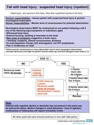
Severity of head injury • mild(13-15 point in Glasgow coma scale) – cerebral concussion, slight cerebral contusion • moderate(8-12 point) – mild cerebral contusion, subacute and chronic cerebral compression • severe(3-7 point)– severe cerebral contusion, diffuse axon injury, acute cerebral compression
Two contusion focuses 1-direct blow on the right 2-countercoup on the left
Linear fractureof occipital bones with going to the skull base
Поперечний зріз аксона, норма • Після травми. відсутні мікротрубочки
Typical location of diffuse axon injury (кружечки) і вогнищ геморагій (заштиховані ділянки)
Cerebral compression • Acute – manifestation during 24 hours afterhead injury • Subacute – manifestation during 1 week afterhead injury • Chronic - manifestation after 1-2 weeks afterhead injury
Causes of cerebral compression • Hematomas • Epidural • Subdural • Intracerebral • Bone fragment at depressed fructures • Pneumocephalus
Main triad at cerebral compression • Deterioration of consciousness level • Ipsilateral anisocoria • contrlateral hemiparesis
Epidural hematoma on the left • Subdural hematoma on the right