Histological and Cytokine Analysis of CCl4-Induced Cirrhotic Liver in Rats
This study presents representative histologic images of CCl4-induced cirrhotic liver using Azan-Mallory staining at x50 magnification. Additionally, cytokine expression levels of IL-1β, IL-8, TNF-α, TGF-β, and RPL21 in the livers of normal and cirrhotic rats were analyzed via RT-PCR. The mRNA expression levels were compared between normal (n=4) and cirrhotic rats (n=4) to explore the inflammatory response associated with liver cirrhosis.

Histological and Cytokine Analysis of CCl4-Induced Cirrhotic Liver in Rats
E N D
Presentation Transcript
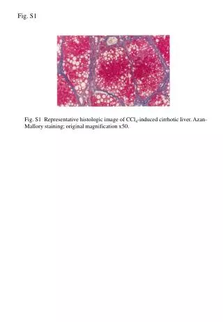
Fig. S1 Fig. S1 Representative histologic image of CCl4-induced cirrhotic liver. Azan-Mallory staining; original magnification x50.
Fig. S2 Cirrhosis Normal IL-1b IL-8 TNF-a TGF-b PRL21 Fig. S2 Cytokine expression levels in the liver of cirrhotic rats. mRNA expression levels of IL-1β, IL-8, TNF-α, TGF-β and RPL21 in the liver of normal rats (n=4) and cirrhotic rats (n=4) were analyzed by RT-PCR using specific primers for each gene. cDNA was synthesized by reverse transcription using total RNA isolated from the liver.


![[Fig. S1]](https://cdn3.slideserve.com/6448662/slide1-dt.jpg)