Impact of TIMP-1 on Vessel Formation and MMP Activity
This study examines the role of TIMP-1 in vessel formation and MMP-dependent processes using fibroblast-conditioned media and siRNA experiments. Results show TIMP-1 enhances vessel formation but is regulated by MMP activity.

Impact of TIMP-1 on Vessel Formation and MMP Activity
E N D
Presentation Transcript
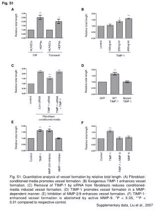
Fig. S1 A B ** Relative total length Relative total length ** * * control 100ng/ml 200ng/ml 400ng/ml HDFNs HUVECs HDFNs HUVECs CM Transwell TIMP-1 C D ** Relative total length Relative total length ** control Con siRNA TIMP-1 siRNA TIMP-1 siRNA + TIMP-1 GFP WT Mutant TIMP-1 TIMP-1 Fibroblast- conditioned media E F ** Relative total length ** Relative total length ** Control TIMP-1 TIMP-1 + MMP-9 MMP-9 Control TIMP-1 MMP-1 inhibitor MMP-2/9 inhibitor Fig. S1. Quantitative analysis of vessel formation by relative total length. (A) Fibroblast-conditioned media promotes vessel formation. (B) Exogenous TIMP-1 enhances vessel formation. (C) Removal of TIMP-1 by siRNA from fibroblasts reduces conditioned-media induced vessel formation. (D) TIMP-1 promotes vessel formation in a MMP-dependent manner. (E) Inhibition of MMP-2/9 enhances vessel formation. (F) TIMP-1-enhanced vessel formation is abolished by active MMP-9. *P < 0.05, **P < 0.01 compared to respective control. Supplementary data, Liu et al., 2007
Fig. S2 A Absorbance 0h 24h 48h B C Con E D TIMP-1 0h 24h Fig. S2. TIMP-1 does not affect proliferation and migration of HUVECs. (A) HUVECs were cultured in a 96-well plate and incubated without (con) or with TIMP-1 (400ng/ml). At indicated time points cell number was determined using methylene blue assay. (B-E) HUVECs were plated in 24-well plate and migration was determined by wound healing assay. The monolayers were wounded and incubated without (con) (B and C) or with TIMP-1 (400ng/ml) (D and E). Supplementary data, Liu et al., 2007
Relative area ** ** con 0.01 0.03 0.1 0.3 1 3 10 MMP-2/9 inhibitor (mM) Fig. S3 Fig. S3. Inhibition of MMP-2/9 enhances vessel formation. HUVECs were grown in three dimensional collagen gel for five days without (con) or with indicated concentrations of MMP-2/9 inhibitor. **P < 0.01 compared to control. Supplementary data, Liu et al., 2007


![[Fig. S1]](https://cdn3.slideserve.com/6448662/slide1-dt.jpg)