Download
1 / 15
150 likes | 519 Vues
Structure of Skeletal Muscle. Presentation by: Angela Holloman. Introduction . All activities that involve movement depend on muscles 650 muscles in the human body Various purposes for muscles for: Locomotion Upright posture Balancing on two legs Support of internal organs

E N D
Structure of Skeletal Muscle Presentation by: Angela Holloman
Introduction • Allactivities that involve movement depend on muscles • 650 muscles in the human body • Various purposes for muscles for: • Locomotion • Upright posture • Balancing on two legs • Support of internal organs • Controlling valves and body openings • Production of heat • Movement of materials along internal tubes • Three types of muscles in the human body • Skeletal • Cardiac • Smooth
Skeletal Muscle • Skeletal muscles are muscles which are attached to the skeleton. • 40% of human body mass • Skeletal muscles are mainly responsible for locomotion, and voluntarycontraction and relaxation.
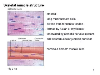
Structure of Skeletal muscles • Skeletal muscles are composed of clusters of muscle cells. • Muscle fibers • Myofibers • Myocytes • A muscle consists of packages of muscle cells called fascicles • A muscle cell is long and spindle shaped
Structure of Skeletal muscles • Cell structure • Muscles cells contain many nuclei • The plasma membrane→ sarcolemma • The cytoplasm→ sarcoplasm • Length • ranges from 0.1cm to more the 30cm in length • Diameter • ranges from 0.001cm to 0.01cm in diameter • Myofibrils→ • elongated protein molecules • aligned in parallel arrangements • extend the full length of the cell.
Structure of Skeletal muscles • The myofibril consists of protein chains called myofilaments • Myofilaments have a symmetrical, alternating pattern of thick and thin elements.
Skeletal Muscle Myosin • Thick myofilament • consists of a large number of bundled myosin molecules aligned in overlapping arrays. • hexameric proteins with two identical heavy chains and two pairs of different light chains. • regulatory light chain (RLC) • essential light chain (ELC)
Skeletal Muscle Actin • The thin myofilament (F-actin, filamentous actin) • made up of two helically intertwined chains of G-actin (globular actin) units. • Other proteins that bind to the actin molecules: • Tropomyosin • The Troponin complex→ made up of three members
Contraction of Skeletal Muscle • The thick and thin filaments, along with their associated myofibril proteins, are responsible for muscle contraction. • How does muscle contraction work? • Influx of calcium ions in the cell • as a result of nerve impulses • troponin complex pulls tropomyosin molecules away from the G-actin subunits • Exposure of the myosin binding sites. • The heads of the myosin molecules can bind to the actin subunits, forming cross bridges. • active site in each myosin head disrupts the high-energy bond of ATP molecules • release of energy moves the myosin head towards the F- actin, • when contact is made with the actin subunits, the F-actin is pulled along, causing the myofilament to contract. • The coordinated contraction of all the myofilaments of all the muscle cells of a muscle, causes the entire muscle to contract.
Relaxation of Skeletal Muscle • Calcium ions are carried away from the myofilaments • Myosin- actin linkages loosen • The troponin complex and tropomyosin bind to the myosin binding sites on the F-actin subunits, • Myosin and F- actin myofilaments return to their original positions
References: • Pasternak, Jack J. Human Molecular Genetics: Mechanisms of Inherited Diseases. Hoboken, New Jersey: John Wilkey & Sons Inc., 2005. • http://www.lau-verlag.de/anatom/muscleb.jpg. March, 20, 2006