Interaction of cIAP1 with TRAF2 & cdc42 Modulates Filopodia Formation
100 likes | 185 Vues
Immunoprecipitation and siRNA analysis reveal cIAP1's interactions with TRAF2 and cdc42, influencing filopodia formation in cells. Silencing cIAPs inhibits TNFα-mediated filopodia induction. cIAP1-deficient cells exhibit increased sensitivity to TNF-induced cell death. HEK293T cells transfected with cIAP1 constructs show cIAP1's ability to interact with cdc42. The study highlights cIAP1's role in modulating cellular filopodia dynamics and cell survival pathways.

Interaction of cIAP1 with TRAF2 & cdc42 Modulates Filopodia Formation
E N D
Presentation Transcript
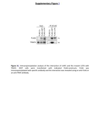
Supplementary Figure 1 FLAG-Co FLAG-cIAP1 FLAG-L47A FLAG-Co FLAG-cIAP1 FLAG-L47A Input IP αFLAG 70 FLAG TRAF2 56 Figure S1. Immunoprecipitation analysis of the interaction of cIAP1 and the mutant L47A with TRAF2. 293T cells were transfected with indicated FLAG-constructs. FLAG was immunoprecipitated with specific antibody and the interaction was revealed using an anti-FLAG or an anti-TRAF antibody.
Supplementary Figure 2 siRNA: control RhoGDIα cIAP1: - + + - cdc42 21 23 70 70 RhoGDIα cIAP1 Figure S2. Immunoblot analysis of cdc42, RhoGDIα and cIAP1 in in NIH3T3 transfected with cIAP1 encoding vector plus Control or RhoGDIα-targeting siRNA. HSC70 is used as loading control. HSC70
53 48 27 Supplementary Figure 3 A IP myc Input EGFP EGFP-Cdc42 EGFP-Rac1 EGFP-RhoA EGFP EGFP-Cdc42 EGFP-Rac1 EGFP-RhoA GFP B GST GST-cIAP1 GTPγS GTPγS GDP GDP lysate NT NT Figure S3. cIAP1 can interact with cdc42. (A) Co-immunoprecipitation analysis of the interaction of cIAP1 with RhoA, Rac1 and cdc42. HEK293T cells were transfected with myc-cIAP1 and EGFP, EGFP-RhoA, EGFP-Rac1 or EGFP-cdc42 encoding vector. Immunoprecipitation was performed using anti-myc antibody and interactions were revealed by immunobloting using anti-GFP antibody. (B) GFP-cdc42 from transfected HEK293T cell lysates was charged with GDP or GTP, and incubated with GST or GST-cIAP1 immobilized onto glutathione sepharose beads. Interactions were revealed by immunoblotting using specific GFP antibody. 48 GFP-Cdc42 GST-cIAP1 95 26
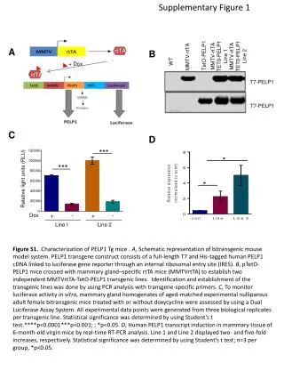
Supplementary Figure 4 * * Filopodia (fold induction/Co) 70 72 70 siRNA: Co cIAP1 cIAP2 cIAP1 cIAP2 HSC 70 siRNA: Co cIAP1 cIAP2 Figure S4. Silencing of cIAPs inhibits TNFα-mediated Filopodia formation. NIH3T3 fibroblasts were transfected with control (Co), cIAP1 or cIAP2 targeting siRNA, serum starved for 16 hours and stimulated for 15 min. with 100ng/mL TNFα. Left panel: The efficiency of siRNA was checked by immunoblot analysis. HSC70 is used as loading control. Right panel: Filopodia were quantified by counting cells exposing more than 5 filopodia. > 100 cells were analyzed. Results were expressed as fold filopodia induction/untreated cells transfected with control siRNA. Mean ± sd of three independent experiments. Statistical analysis performed using the Student’s t-test. *: p<0,05.
Supplementary Figure 5 wt cIAP1-/- NT • 125 • 100 • 75 • 50 • 25 • 00 Survival (% ) TNF 4h TNF 8h Figure S5. MEF cIAP1-/- are sensitive to TNF-induced cell death. MEF wt or cIAP1-/-were serum starved for 16 hours, then stimulated for 4 or 8 hrs with 100ng/mL TNF. Cell viability was evaluated by a crystal violet staining (Left panel: One representative experiment is shown) and quantified after elution by a colorimetric analysis (Right panel: Mean ± sd of three independent experiments).
Supplementary Figure 6 GFP-Co GFP-L47A GFP-cIAP1 GFP-Co GFP-L47A GFP-cIAP1 Input IP αGFP cIAP1 GFP 96 70 GFP-cIAP1/L47A Endogenous cIAP1 96 70 Lower exposure GFP-cIAP1/L47A Heavy chains GFP 96 26 Figure S6. HEK293T cells were transfected with GFP, GFP-cIAP1 or GFP-cIAP1L47A (GFP-L47A). GFP-IAP constructs were Immunoprecipitated using anti-GFP antibody and interactions were revealed by immunobloting using anti-cIAP1 antibody detecting the overexpressed constructs and endogenous cIAP1.
Supplementary Figure 7 *** ** ** *** Filopodia (fold inductin / UT) si-Co si-TRAF2 Co Co cIAP1 MEFs: wt cIAP1-/- wt Figure S7. MEFs from wt or cIAP1-/- mice were transfected or silenced as in Figure 5C-E, serum starved, and treated for 10 minutes with 100ng/mL EGF. Filopodia were evaluated by a Microscopic analysis of F-actin immunostained using AlexaFluor488-conjugated Phalloidin and counted. > 100 cells were analyzed. Mean ± sd of three independent experiments. Statistical analysis performed using the Student’s t-test. ***: p<0,001; **: p<0,01; *: p<0,05.
Supplementary Figure 8 Figure S8. Nude mice were injected in the tail vein wih 1.106 MEF wt or cIAP1-/- expressing Hras-V12 and the mice were sacrificed 2 weeks later. Pictures of the whole lungs (upper panel) or lung sections stained with hematoxylin and eosin (lower panel) are presented.
Supplementary Figure 9 Figure S9. CFSE-labeled wt MEFs expressing HRas-V12 were added onto confluent HUVECs and filmed for 300 min. by time-lapse microscopy (picture every 15 min.) The intercalated cells that display a non-round shape and a low phase-bright were marked with an Asterisk. The arrow indicates the cell that did not interacalate
Supplementary Figure 10 cIAP1: - - - + - - + His-Ub: + - + + - + + HA-cdc42: - wt wt wt L61 L61 L61 130 Co2+ Pull down HA (ubiquitin) 72 43 26 cIAP1 72 HA 26 Figure S10. HEK293T were transfected with His-ubiquitin, HA-cdc42, HA-cdc42L61 (L61) constitutive active mutant of cdc42 and cIAP1. His-ubiquitin was precipitated by using cobalt beads and ubiquitinated proteins were revealed by immunobloting by using anti-HA specific antibody. The efficiency of transfection was checked by immunobloting by using anti-cIAP1 and anti-HA antibodies.