Download
1 / 15
150 likes | 178 Vues
Explore advanced neuroimaging techniques for detecting mild traumatic brain injuries with implications for improved diagnostic accuracy and treatment outcomes. This research aims to bridge the gap in translating neuroimaging findings into clinical practice.

E N D
Neuroimaging Detection of mTBI Abnormalities Christine Mac Donald, PhD Assistant Professor Department of Neurological Surgery University of Washington School of Medicine Seattle, WA USA
Motivation for Research • 1. Coronado VG, McGuire LC, Sarmiento K, et al. Trends in Traumatic Brain Injury in the U.S. and the public health response: 1995-2009. Journal of safety research. 2012;43(4):299-307. • Centers for Disease Control and Prevention (CDC) NCfIPaC. Report to Congress on mild traumatic brain injury in the United States: steps to prevent a serious public health problem. Atlanta (GA): Centers for Disease Control and Prevention2003. • Kashluba S, Hanks RA, Casey JE, Millis SR. Neuropsychologic and functional outcome after complicated mild traumatic brain injury. Arch Phys Med Rehabil. 2008;89:904-11. • Iverson GL. Mild traumatic brain injury meta-analyses can obscure individual differences. Brain Inj. 2010;24:1246-55. • Lee H, Wintermark M, Gean AD, Ghajar J, Manley GT, Mukherjee P. Focal lesions in acute mild traumatic brain injury and neurocognitive outcome: CT versus 3T MRI. J Neurotrauma. 2008;25(9):1049-56. • Gentry LR, Godersky JC, Thompson B, Dunn VD. Prospective comparative study of intermediate-field MR and CT in the evaluation of closed head trauma. AJR Am J Roentgenol. 1988;150:673-82. • Jenkins A, Hadley MDM, Teasdale G, Macpherson P, Rowan JO. Brain lesions detected by magnetic resonance imaging in mild and severe head injuries. Lancet. 1986;2(8504):445-6. • Mittl RL, Grossman RI, Hiehle JF, et al. Prevalence of MR evidence of diffuse axonal injury in patients with mild head injury and normal head CT findings. AJNR Am J Neuroradiol. 1994;15:1583-9. • Orrison WW, Gentry LL, Stimac GK, Tarrel RM, Espinosa MC, Cobb LC. Blinded comparison of cranial CT and MRI in closed head injury evaluation. AJNR Am J Neuroradiol. 1994;15:351-6. • Traumatic brain injury (TBI) affects approximately 3.5 million individuals annually in the United States1 and approximately 75% are due to ‘mild’ or concussive events2. • Current clinical and diagnostic tools for assessing concussion are insensitive • Clinical – Glasgow Coma Score - has major limitations obscuring differences among diverse subgroups of TBI patients with very different prognoses3,4. • Diagnostic –CT – Currently only modality recognized by the FDA but well established that head CT is grossly insensitive to concussion pathology. • An objective measure of concussion is lacking • Multi-modal Magnetic Resonance Imaging (MRI) has diagnostic appeal but has yet to be validated for regulatory readiness as a screening tool for concussion. • Prior work has shown that MRI has much greater sensitivity than CT for small, focal traumatic intracranial lesions following brain injury5-9 but what about concussion pathology as a whole?
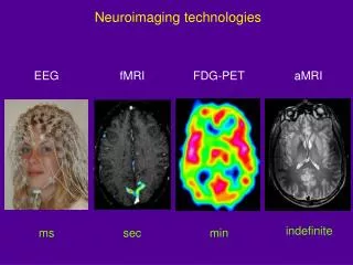
MR Imaging of Concussion Resting-State functional MRI and fMRI Methods2 Diffusion Tensor Imaging and Higher Order Models High Resolution Structural Volumetric Imaging And So On… Prior work has shown that approximately one-third of patients presenting to the ED with head injury, but with no head CT findings, have acute trauma-related brain pathology on conventional MRI1qualitatively interpreted by a Radiologist. These findings have been correlated to clinical and functional outcome following concussion1. Research with Advanced MR methods has also provided further insight but inconsistencies have been reported in approach, analysis, and findings. Not just ‘pretty pictures’ – quantitatively informative results Yuh EL, Mukherjee P, Lingsma HF, Yue JK, Ferguson AR, Gordon WA, Valadka AB, Schnyer DM, Okonkwo DO, Maas AI, Manley GT; TRACK-TBI Investigators. Magnetic resonance imaging improves 3-month outcome prediction in mild traumatic brain injury. Ann Neurol. 2013 Feb;73(2):224-35. doi: 10.1002/ana.23783. Epub 2012 Dec 7. Zhou Y, et al "Default-mode network disruption in mild traumatic brain injury" Radiology 2012; 265: 882-892.
The Challenge Many were single site studies with no validation across MRI platforms Group level differences were often reported without showing sensitivity on a single-subject level For quantitative methods, there had yet to be standardization in acquisition protocol, post-processing pipelines or analytical approach Little consideration had been given to how to directly compare quantitative data collected from different machines when systematic differences in hardware have been shown to impact these results with varying degrees of significance With millions of grant dollars spent, we have yet to successfully translate any of these methods into clinical practice reaching our end goal of: Providing new diagnostic biomarkers for concussion/brain injury
New Avenues Towards Successful Translation Pushing the Field Forward Current Achievements from TRACK-TBI A ‘Big Data’ Approach to Resolving the ‘Small Study’ Problem
How TRACK-TBI Differs from Prior Work • 11 site multi-center study working across the G-P-S system of vendors • GE: UCSF, U Cincinnati, UT-Southwestern, U Pitt • Phillips: UW, UT-Southwestern, VCU • Siemens: U Maryland, Harvard/MGH, UCSF, U Pitt, UT Austin, U Miami, VCU, Baylor • First major TBI study to provide a standardized protocol across all platforms for imaging acquisition which includes advanced MRI methods • One of few to complete site qualification and monthly quality assurance methods utilizing three Imaging Phantoms for quantitative combination of data across imaging platforms • ADNI Phantom – Structural Imaging • BIRN Phantom – BOLD (fMRI) Imaging • HPD Phantom – Diffusion Imaging • Providing supportive evidence for feasibility of MR imaging across age spectrum (0-100 years) and injury severity (mild-moderate-severe) • Prior efforts have selected strict inclusion criteria however more often than not, there is high co-morbidity to other disease states with brain injury
HPD (Diffusion) ADNI (Structural) BIRN (fMRI) Providing a ‘ground truth’ from which to align data
TRACK – TBI Imaging Progress Queried 22 May 2015
New Challenges arising from New Approach Diffusion Tensor Phantom • Data sharing and dissemination • Providing a central repository for ‘big’ imaging data • Average scan 3-500MB, 1000 scans ~ 500TB • Processed data ~2-20TB, 1000 scans ~2-20,000TB! (20 petabytes) • More than what can be handled comfortably on a cloud really at the level of industry multi-server storage • Expedited data transfer from around the world • Supportive evidence from imaging applications in combat • Further development of phantoms and calibration algorithms required for data alignment • Diffusion – Diffusivity to Anisotropy or Hybrid of both • Utilization of current data to optimize post-processing pipelines Farrher E, Kaffanke J, Celik AA, Stöcker T, Grinberg F, Shah NJ. Novel multisection design of anisotropic diffusion phantoms. MagnReson Imaging. 2012 May;30(4):518-26
Mild Brain Injury Patient (Same Data -Different Pipelines) Pipeline A Pipeline B *Images have been windowed slightly overexposed to highlight discrepancies
Severe Brain Injury Patient (Same Data -Different Pipelines) Pipeline C Pipeline B
Moving Forward http://www.cloudcomputing-news.net/media/img/news/iStock_2015_2.jpg.800x600_q96.png
Next Steps • Show clinical utility across all imaging platforms • Dissemination to the masses • ‘Big data’ studies for validation (TRACK-TBI, CENTER-TBI, etc) • Explore opportunities for further collaboration and combination of existing data • Design and refinement of phantoms, calibration algorithms • Standardization of quantitative post-processing pipelines • Direct application and translation of imaging methods to other neurodegenerative disease states • Parkinson's • Alzheimer's Disease • ALS • Multiple Sclerosis • ……
Successful Translation - End Goals • Provide more sensitive tools for: • Diagnosis • Rehabilitation • Stratification for Therapeutic intervention/Drug development • Reconsideration to previously ‘failed’ drug trials? • Were previous results influenced by crude stratification measures? • CT scan negative for control may not have been a control (?) • Lower GCS for TBI may have been for other concomitant medical issues not a real TBI (?) • Remember: How will this help the patient • Provide more definitive evidence for disability claims • Who is really in need and who is just looking for $$$ • Provide further piece of mind about underlying condition even in the absence of injury resolution
Thank you! Follow up comments/questions can be directed to: cmacd@uw.edu