Download
1 / 80
850 likes | 1.37k Vues
Principles of Neuroimaging . Michael S. Beauchamp, Ph.D. Assistant Professor Department of Neurobiology and Anatomy. Michael.S.Beauchamp@uth.tmc.edu. Friday, February 25th@ 8 a.m., MSB 2.006 M1 Medical Neuroscience. Nachum Dafny , Course Director. Why is neuroimaging difficult?. 2 - 17.

E N D
Principles of Neuroimaging Michael S. Beauchamp, Ph.D. Assistant Professor Department of Neurobiology and Anatomy Michael.S.Beauchamp@uth.tmc.edu Friday, February 25th@ 8 a.m., MSB 2.006 M1 Medical Neuroscience. NachumDafny, Course Director
Why is neuroimaging difficult? 2 - 17 17 - 21 21 - 23
Palpation/Sensation • skull: can’t feel much from outside (sorry, ultrasound) • -no nerves in brain: can’t feel much from inside
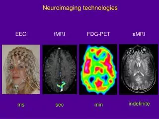
Alphabet Soup SPECT CT MRI PET MEG EEG fMRI
Accessory (non-imaging) Methods Neuroimaging Methods External Ionizing Radiation MEG EEG Internal Ionizing Radiation Non-ionizing radiation (Radio Waves) CT PET MRI Angiogram/ Arteriogram SPECT fMRI MR Angiography X-Ray (radiograph) Diffusion Imaging (DWI/DTI) MR Spectroscopy Taxonomy of Neuroimaging Methods
Accessory (non-imaging) Methods Neuroimaging Methods External Ionizing Radiation MEG EEG Internal Ionizing Radiation Non-ionizing radiation (Radio Waves) CT PET MRI Angiogram/ Arteriogram SPECT fMRI MR Angiography X-Ray (radiograph) Diffusion Imaging (DWI/DTI) MR Spectroscopy Taxonomy of Neuroimaging Methods
Accessory (non-imaging) Methods Neuroimaging Methods External Ionizing Radiation MEG EEG Internal Ionizing Radiation Non-ionizing radiation (Radio Waves) CT PET MRI Angiogram/ Arteriogram SPECT fMRI MR Angiography X-Ray (radiograph) Diffusion Imaging (DWI/DTI) MR Spectroscopy Taxonomy of Neuroimaging Methods
Accessory (non-imaging) Methods Neuroimaging Methods External Ionizing Radiation MEG EEG Internal Ionizing Radiation Non-ionizing radiation (Radio Waves) CT PET MRI Angiogram/ Arteriogram SPECT fMRI MR Angiography X-Ray (radiograph) Diffusion Imaging (DWI/DTI) MR Spectroscopy Taxonomy of Neuroimaging Methods
Arteriogram (a.k.a. Angiogram) Basic principle: Inject contrast agent (dye) that is radio-opaque i.e. iodine containing agents
Accessory (non-imaging) Methods Neuroimaging Methods External Ionizing Radiation MEG EEG Internal Ionizing Radiation Non-ionizing radiation (Radio Waves) CT PET MRI Angiogram/ Arteriogram SPECT fMRI MR Angiography X-Ray (radiograph) Diffusion Imaging (DWI/DTI) MR Spectroscopy Taxonomy of Neuroimaging Methods
CT (computed tomography) Pros: Widely available Very fast to collect whole-head images (one slice in < 1 ms; whole head in ~ seconds; whole exam in 10 minutes ) Somewhat cheaper than MRI (~$500 vs ~$1000 for MRI) Less hassle (few contraindications) Best for an emergency Cons: Exposure to ionizing radiation (increased risk of cancer) can require a contrast agent—for brain, injectable iodine compound Poor tissue contrast not versatile
CT (computed tomography) Basic Principle: rotate machinery to take multiple x-rays with different paths through the body
Common clinical use: stroke Patient presents with stroke 80% 20% ischemic hemorraghic Give tPA, dissolve clot, Blood flow restored, Patient recovers Give tPA, prevent clotting, Patient dies of massive bleed
Common clinical use: stroke Patient presents with stroke 80% 20% ischemic hemorraghic
CT (computed tomography) Pros: Widely available Very fast to collect whole-head images (one slice in < 1 ms; whole head in ~ seconds; whole exam in 10 minutes ) Somewhat cheaper than MRI (~$500 vs ~$1000 for MRI) Less hassle (few contraindications) Best for an emergency Cons: Exposure to ionizing radiation (increased risk of cancer) can require a contrast agent—for brain, injectable iodine compound Poor tissue contrast not versatile
Accessory (non-imaging) Methods Neuroimaging Methods External Ionizing Radiation MEG EEG Internal Ionizing Radiation Non-ionizing radiation (Radio Waves) CT PET MRI Angiogram/ Arteriogram SPECT fMRI MR Angiography X-Ray (radiograph) Diffusion Imaging (DWI/DTI) MR Spectroscopy
Accessory (non-imaging) Methods Neuroimaging Methods External Ionizing Radiation MEG EEG Internal Ionizing Radiation Non-ionizing radiation (Radio Waves) CT PET MRI Angiogram/ Arteriogram SPECT fMRI MR Angiography X-Ray (radiograph) Diffusion Imaging (DWI/DTI) MR Spectroscopy “Nuclear Medicine”
“Nuclear Medicine”: PET/SPECT Basic priniciple: Inject radioactive isotope attached to metabolic compound (Oxygen, Glucose, etc.) Wait for it to decay, look for radioactive decay products
PET/SPECT Pros: Fairly cheap (~ $1000) Shows function (metabolism) Cons: Exposure to ionizing radiation (increased risk of cancer) ~1 year of background radiation Low-resolution Slow to very slow not versatile
SPECTSingle Photon Emission Computed Tomography “Nuclear Medicine”:~unclear medicine
SPECTSingle Photon Emission Computed Tomography • Radionuclides • Single-Gamma emitting • 99mTc, 123I, 67Ga, 111In
PET: Positron Emission Tomography Basic priniciple: Inject radioactive isotope attached to important metabolic compound (Oxygen, Glucose) Wait for it to decay. Pick up two particles going in opposite directions—improves spatial resolution • PET uses beta-plus-emitting radionuclides such as C-11, N-13, O-15, and F-18 which annihilate into two 511-keV photons that travel in opposite directions.
Developments in PET [11C] Flumazenil (benzodiazepine receptor antagonist ~ GABA-A ) [11C]PMP Substrate for AChE [11C]DTBZ VMAT2 ~dopamine,serotonin • Development of new radiotracers Joshi et al., JCBFM 2009
Sample Clinical Application: Alzheimer’s Diagnosis • Compounds that bind to AD plaques • Conditional approval by FDA (Jan 2011) • C. M. Clark et al. J. Am. Med. Assoc. 305, 275–283; 2011
Accessory (non-imaging) Methods Neuroimaging Methods External Ionizing Radiation MEG EEG Internal Ionizing Radiation Non-ionizing radiation (Radio Waves) CT PET MRI Angiogram/ Arteriogram SPECT fMRI MR Angiography X-Ray (radiograph) Diffusion Imaging (DWI/DTI) MR Spectroscopy
MRI: Magnetic Resonance Imaging Pros: incredible images advancing very rapidly extremely high resolution extremely versatile NO ionizing radiation Cons: moderately expensive (~$1000) complex some contraindications Can require injection of contrast agents (Gadolinium/Iron compounds)
Imaging Techniques: MRI • the MR scanner is a giant magnet: 1.5T, 3T, 7T, 9T
Contraindications I • Ferrous metal in body
Contraindications II • Cochlear implants (always) • Maybe: pacemakers, vagal nerve stimulators, old (> 20 year) surgical implants
UTH MRI Facility Location: Ground Floor, MSB Building Equipment: Phillips 3T whole body human MRI scanner Mock scanner for testing and training Bruker 7T small animal (rodent) scanner
MRI (Magnetic Resonance Imaging) Basic principle: uses radio waves to interrogate protons in water molecules in the brain
MRI (Magnetic Resonance Imaging) Basic principle: uses radio waves to interrogate protons in water molecules in the brain
We listen to these radio waves with an “RF coil” (radio antenna) H2O H2O H2O
[Main magnet and some of its lines of force] [Little magnets lining up with external lines of force] B0 = Giant Field Produced by Giant Magnet • Purpose is to align H protons in H2O (little magnets)
Small B0 produces • small net • magnetization M • Thermal motions try to randomize alignment of proton magnets • Larger B0 produces • larger net • magnetization M, • lined up with B0 • Reality check: 0.0003% of protons • aligned per Tesla • of B0
Precession of Magnetization M • Magnetic field causesMto rotate (or precess) about the direction of B at a frequency proportional to the size of B — 42 million times per second (42 MHz), per Tesla of B • If M is not parallel to B, then • it precesses clockwise around • the direction of B. • However, “normal” (fully relaxed)situation has M parallel to B, which means there won’t be any • precession • N.B.: part of M parallel to B (Mz) does not precess
B1 = Excitation (Transmitted) Radio Frequency (RF) Field • Left alone, M will align itself with B in about 2–3 s • So don’t leave it alone: apply (transmit) a magnetic field B1 that fluctuates at the precession frequency and points perpendicular to B0 • The effect of the tiny B1is to cause M to spiral away • from the direction of the • static B field • B110–4 Tesla • This is called resonance • If B1 frequency is not close to resonance, B1has no effect Time = 2–4 ms
Relaxation: Nothing Lasts Forever • In absence of external B1, M will go back to being aligned with static field B0— this is called relaxation • T2: Part of M perpendicular to B0shrinks [Mxy] • This part of M is called transverse magnetization • It provides the detectable RF signal • T1: Part of Mparallel to B0 grows back [Mz] • This part of M is called longitudinal magnetization • Not directly detectable, but is converted into transverse magnetization by externally applied B1
Basics of MR • T1 ~ Longitudinal Magnetization/Relaxation “hi-resolution, normal anatomy” T2 ~ Transverse Magnetization/Relaxation “pathology”—water content
Basics of MR—contrast agents • Gadolinium • can be injected to enhance contrast (usually in T1 images); hastens T1 recovery making image brighter
Clinical Applications: Multiple Sclerosis • T2 is best for seeing white matter abnormalities