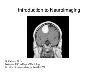
Introduction to Neuroimaging
750 likes | 1.25k Vues
Introduction to Neuroimaging. Aaron S. Field, MD, PhD Assistant Professor of Radiology Neuroradiology Section University of Wisconsin–Madison. Updated 7/17/07. Neuroimaging Modalities. Magnetic Resonance (MR) MR Angiography/Venography (MRA/MRV) Diffusion and Diffusion Tensor MR

Introduction to Neuroimaging
E N D
Presentation Transcript
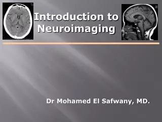
Introduction to Neuroimaging Aaron S. Field, MD, PhD Assistant Professor of Radiology Neuroradiology Section University of Wisconsin–Madison Updated 7/17/07
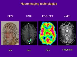
Neuroimaging Modalities • Magnetic Resonance (MR) • MR Angiography/Venography (MRA/MRV) • Diffusion and Diffusion Tensor MR • Perfusion MR • MR Spectroscopy (MRS) • Functional MR (fMRI) • Nuclear Medicine • Single Photon Emission Computed Tomography (SPECT) • Positron Emission Tomography (PET) • Radiography (X-Ray) • Fluoroscopy (guided procedures) • Angiography • Diagnostic • Interventional • Myelography • Ultrasound (US) • Gray-Scale • Color Doppler • Computed Tomography (CT) • CT Angiography (CTA) • Perfusion CT • CT Myelography “Duplex”
Radiography (X-Ray) • Primarily used for spine: • Trauma • Degenerative Dz • Post-op
Fluoroscopy (Real-Time X-Ray) • Fluoro-guided procedures: • Angiography • Myelography
Fluoroscopy (Real-Time X-Ray) Digital Subtraction Angiography
Fluoroscopy (Real-Time X-Ray) Digital Subtraction Angiography
Digital Subtraction Angiography Indications: • Aneurysms, vascular malformations and fistulae • Vessel stenosis, thrombosis, dissection, pseudoaneurysm • Stenting, embolization, thrombolysis (mechanical and pharmacologic) • Ability to intervene • Time-resolved blood flow dynamics (arterial, capillary, venous phases) • High spatial and temporal resolution • Invasive, risk of vascular injury and stroke • Iodinated contrast and ionizing radiation Advantages: Disadvantages:
Fluoroscopy (Real-Time X-Ray) Myelography Lumbar or cervical puncture Inject contrast intrathecally with fluoroscopic guidance Follow-up with post-myelo CT (CT myelogram)
Myelography Indications: • Spinal stenosis, nerve root compression • CSF leak • MRI inadequate or contraindicated • Defines extent of subarachnoid space, identifies spinal block • Invasive, complications (CSF leak, headache, contrast reaction, etc.) • Ionizing radiation and iodinated contrast • Limited coverage Advantages: Disadvantages:
US transducer carotid Ultrasound
Ultrasound Indications: • Carotid stenosis • Vasospasm - Transcranial Doppler (TCD) • Infant brain imaging (open fontanelle = acoustic window) • Noninvasive, well-tolerated, readily available, low cost • Quantitates blood velocity • Reveals morphology (stability) of atheromatous plaques • Severe stenosis may appear occluded • Limited coverage, difficult through air/bone • Operator dependent Advantages: Disadvantages:
Ultrasound – Gray Scale Gray-scale image of carotid artery
Ultrasound – Gray Scale Plaque in ICA Gray-scale image of carotid artery
Ultrasound - Color Doppler Peak Systolic Velocity (cm/sec)ICA Stenosis (% diameter) 125 – 225 50 – 70 225 – 350 70 – 90 >350 >90
Computed Tomography A CT image is a pixel-by-pixel map ofX-ray beam attenuation(essentiallydensity)inHounsfield Units (HU) HUwater = 0 Bright = “hyper-attenuating” or “hyper-dense”
Computed Tomography Typical HU Values: Air –1000 Fat –100 to –40 Water 0 Other fluids (e.g. CSF) 0–20 White matter 20–35 Gray matter 30–40 Blood clot 55–75 Calcification >150 Bone 1000 Metallic foreign body >1000 Brain
Computed Tomography Attenuation: High or Low? • High: • Blood, calcium • Less fluid / more tissue • Low: • Fat, air • More fluid / less tissue Air –1000 Fat –100 to –40 Water 0 Other fluids 0–20 White matter 20–35 Gray matter 30–40 Blood clot 55–75 Calcification >150 Bone 1000 Metallic foreign body >1000
Computed Tomography “Soft Tissue Window” “Bone Window”
Computed Tomography Scan axially… …stack and re-slice in any plane “2D Recons”
CT Indications • Skull and skull base, vertebrae • (trauma, bone lesions) • Ventricles • (hydrocephalus, shunt placement) • Intracranial masses, mass effects • (headache, N/V, visual symptoms, etc.) • Hemorrhage, ischemia • (stroke, mental status change) • Calcification • (lesion characterization)
Skull and skull base, vertebrae Fractures
Skull and skull base, vertebrae Multiple Myeloma Osteoma
Ventricles Hydrocephalus
Intracranial masses, mass effects Solid mass Cystic mass
Intracranial masses, mass effects L hemisphere swelling Generalized swelling
Acute Hemorrhage Intraparenchymal Subarachnoid Subdural Epidural
Acute Ischemia Loss of gray-white distinction and swelling in known arterial territory
Calcification Hyperparathyroidism
CT Angiography • Rapid IV contrast bolus • Dynamic scanning during arterial phase • Advanced 2D and 3D Reconstructions: • 2D multi-planar (sagittal, coronal) • Volume–rendered 3D recons
CT Angiography - Head Circle of Willis Vascular Malformations Aneurysms
CT Angiography - Neck Carotid bifurcations Vertebral arteries Aortic arch
CT Angiography 3D Volume Rendering
CT Angiography - Indications • Atherosclerosis • Thromboembolism • Vascular dissection • Aneurysms • Vascular malformations • Penetrating trauma
CBV CBF MTT CT Perfusion
Hemodynamic Parameters Derived From Concentration-Time Curves Bolus arrival Vein Artery
Hemodynamic Parameter Maps Transit Time (sec) Blood Flow (mL/min/g) Blood Volume (mL/g)
CT Myelography • Spinal CT immediately following conventional myelogram • Cross-sectional view of spinal canal along with spinal cord and nerve roots • Assess spinal stenosis/nerve root compression (e.g. disc herniation, vertebral fracture, neoplasm)
Magnetic Resonance (MR) Hydrogen proton in water or fat MRI
Magnetic Resonance Imaging Transmitter Receiver RF COMPUTER RF = Radio Frequency energy Received signal magnetic field
Magnetic Resonance Safety MRI Safety Test: Will it: Move? Torque? Get hot? Pass a current? Malfunction? Become a projectile? Get stuck in scanner? • Typically unsafe*: • Cardiac pacemakers (and other electrical devices) • Some older aneurysm clips • Metal fragments in orbit (1 case report) • Oxygen tanks, carts, chairs, stools, IV poles, gurneys, etc. • Some cosmetics, tattoos, jewelry, hairpins, etc. • Pager, watch, wallet, ID badge, pen, keys, pocketknife, etc. • Typically safe*: • Orthopedic hardware • Surgical clips, staples, sutures (older devices must be checked!) • Intravascular stents/filters * This is an incomplete list and there are many exceptions to every “rule” When in doubt, check it out!