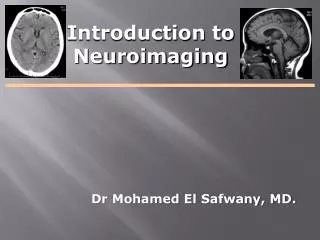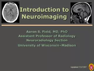
Introduction to Neuroimaging
1.18k likes | 1.51k Vues
Dive into detailed insights on spinal imaging techniques, anatomy, common pathologies including trauma, degenerative diseases, tumors, and more. Learn to evaluate fractures, ligamentous injuries, and degenerative disc conditions with imaging modalities like MRI and CT scans.

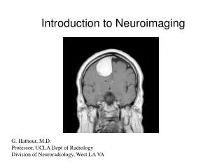
Introduction to Neuroimaging
E N D
Presentation Transcript
Introduction to Neuroimaging SPINE Ayşegül Sarsılmaz, MD RadiologyDepartment Yeditepe University
Spine Pathology • Trauma • Degenerative disease • Tumors and other masses • Inflammation and infection • Vascular disorders • Congenital anomalies
Evaluating Trauma • Fracture – plain film / CT • Dislocation – plain film / CT • Ligamentous injury – MRI • Cord injury – MRI • Nerve root avulsion – MRI
Plain film findings may be very subtle or absent! Anterolisthesis of C6 on C7 (Why??)
CT Fractures of C6 left pedicle and lamina
CT – 2D Reconstructions Acquire images axially… …reconstruct sagittal / coronal
Vertebral body burst fx with retropulsion into spinal canal 2D Reformats
Vertebral Artery Dissection/Occlusion Secondary to C6 Fracture
Hyperflexion fx with ligamentous disruption and cord contusion
Nerve root avulsion Axial Coronal Sagittal
Degenerative Disc (and Facet Joint) Disease Foraminal stenosis Thickening/Buckling of Ligamentum Flavum
Lumbar Spinal Stenosis Disc bulge, facet hypertrophy and flaval ligament thickening frequently combine to cause central spinal stenosis Note the trefoil shape of stenotic spinal canal
Lumbar Spinal Stenosis Disc bulge, facet hypertrophy and flaval ligament thickening frequently combine to cause central spinal stenosis Note the trefoil shape of stenotic spinal canal
Neural foramen Foraminal Stenosis