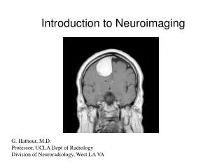
NeuroImaging
NeuroImaging. Dr. Norman Pay. CT. Transmission. CT. Transmission Density differences Ionizing radiation Iodinated contrast material Spatial resolution Fast scanning times and acquisition Appropriate in emergent situations, claustrophobic patients, body coverage


NeuroImaging
E N D
Presentation Transcript
NeuroImaging Dr. Norman Pay
CT • Transmission
CT • Transmission • Density differences • Ionizing radiation • Iodinated contrast material • Spatial resolution • Fast scanning times and acquisition • Appropriate in emergent situations, claustrophobic patients, body coverage • Utilization for contraindications in MRI as aneurysm clips, cardiac pacemakers, etc. • Biopsies • Workstation compatibility • CT angiography
RADIATION • Sv (Sievert) – absorbed dose in biological tissue • 2 mSv/ year – background radiation • 24 mSv/ year –background radiation for airline cruising altitude • 6.8 mSv – chest CT scan • 10-30 mSv – single full body CT scan • 21 Sv – fatal dose
CT Angiography CAROTID TRAUMATIC ANEURYSM CAROTID STENT
MRI • Proton Relaxation • Signal intensities • Contrast resolution • Gadolinium (Gd) contrast • Nephrogenic Systemic Fibrosis (NSF) – Gd contraindicated in Low GFR states (<30) and renal failure • Non-ionizing, non-invasive • Workstation compatibility • More complex, longer acquisitions and set-up • Magnet bore - claustrophobia • MR angiography
MR Angiography CAVERNOMA CAROTID DISSECTION CAROTID OCCLUSION
T2 Flair T1 GRE Diffusion Contrast
MR sequences • T1 – anatomy, CSF dark • T2 – screening, CSF bright • FLAIR (fluid attenuated inversion recovery) – similar to T2 • MR diffusion – bright signal for restriction • GRE (gradient echo) – susceptibility- dark signal • Gadolinium, T1 – bright signal • MR angiography and perfusion – Gadolinium utilization
Anatomy of the Brain • Spatial Resolution • CT Density • Contrast Resolution • MR Signal Intensity • Intravenous Contrast • Iodinated contrast • Gadolinium contrast
T2 FLAIR CORTICAL DYSPLASIA
Stroke • Acute ischemic stroke (AIS) – 3rd leading cause of death, leading cause of disability in adults • 700,000 ischemic strokes annually in the U.S. • Reperfusion therapy is the only proven treatment of AIS
CT and MR • Time to infarct • Time to treatment • Extent of infarct • Hematoma • Recovery
POST THROMBUS LYSIS PRE THROMBUS LYSIS
CT POST THROMBUS LYSIS PRE THROMBUS LYSIS
MOYA-MOYA FLAIR
Pattern Recognition Diagnostic Neuroradiology, pg 130-131. Osborn, Anne G., M.D. Mosby – Year Book, Inc., 1994.
Pattern Recognition Diagnostic Neuroradiology, pg 130-131. Osborn, Anne G., M.D. Mosby – Year Book, Inc., 1994.
MR DIFFUSION • Diffusion refers to the general transport of molecules, mixing through agitation and randomly • The driving force is the motion of water within water, driven by thermal agitation called Brownian motion • If restricted as in acute infarcts, decreased diffusion results • Decreased diffusion displayed as bright MR signal
MR DIFFUSION • Failure of Na+/K+ ATPase and other ionic pumps – net shift of water from the extracellular to the intracellular space • Cell swelling with decrease in extracellular space • Increased intracellular viscosity and cell membrane permeability • Temperature decrease • Decreased diffusion in acute stroke
CEREBELLAR INFARCT CT MR DIFFUSION
CT MR MIDDLE CEREBRAL ARTERY INFARCT
DIFFUSION DIFFUSION DIFFUSION MRA MRA BASILAR ARTERY OCCLUSION
DIFFUSION FLAIR ACUTE INFARCT
MR DIFFUSION EMBOLIC DISEASE – ATRIAL FIBRILLATION
T1 T2 T1 CONTRAST POSTERIOR CEREBRAL ARTERY INFARCT
FLAIR T2 FLAIR VASCULITIS
DIFFUSION DIFFUSION FLAIR FLAIR STATUS POST AORTIC VALVE SURGERY HYPOTENSION
Neuroimaging in acute ischaemic stroke: insights into unanswered questions of pathophysiology. Wardlaw, J. M. Journal of Internal Medicine 267; 172–190. Blackwell Publishing Ltd. 2010.
MR DIFFUSION Neuroimaging in acute ischaemic stroke: insights into unanswered questions of pathophysiology. Wardlaw, J. M. Journal of Internal Medicine 267; 172–190. Blackwell Publishing Ltd. 2010.
CT MR MIDDLE CEREBRAL ARTERY INFARCT
Hematoma • Hemorrhagic transformation – dreaded complication • Exclusion of hematoma -prerequisite for treatment • Cue for emergent intervention
HEMORRHAGE INTO INFARCT INFARCT CT
HEMATOMA EPIDURAL EPIDURAL SUBDURAL
REBLEED ISODENSE SUBDURAL HEMATOMA
Magnetic Resonance Imaging of the Brain and Spine, 3rd ed., Vol. 1, pg 788. Atlas, Scott W., M.D., editor. Lippincott Williams & Wilkins, 2002.
FLAIR GRE HEMATOMA
T1 T2 CT SUBDURAL HYGROMA AND HEMATOMA
T1 CT T2 CHRONIC CHRONIC CHRONIC SUBDURAL HEMATOMA
FLAIR FLAIR CT SUBARACHNOID HEMORRHAGE
T1 FLAIR T1 VENOUS THROMBOSIS AND VENOUS INFARCT
GRE SIDEROSIS
HEMATOMA FLAIR CT GRE MALIGNANT MALIGNANT BENIGN
T1 T2 CONTRAST MALIGNANT HEMATOMA
T1 GRE CT T2 CYST
SUMMARY • CT and MR utilize different technologies, often complementary • Advantages and disadvantages of CT and MR • CT and MR advances pari-passu with computing capabilities • Moore’s Law
REFERENCES • Diagnostic Neuroradiology, pg 130-131. Osborn, Anne G., M.D. Mosby – Year Book, Inc., 1994. • Magnetic Resonance Imaging of the Brain and Spine, 3rd ed., Vol. 1, pg 788. Atlas, Scott W., M.D., editor. Lippincott Williams & Wilkins, 2002. • Neuroimaging in acute ischaemic stroke: insights into unanswered questions of pathophysiology. Wardlaw, J. M. Journal of Internal Medicine 267; 172–190. Blackwell Publishing Ltd. 2010.